ChIP-seq Decoded: Mapping Histone Modifications and Non-Coding RNAs in Disease Research and Drug Discovery
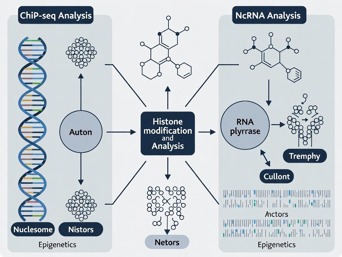
This article provides a comprehensive guide for researchers and drug development professionals on applying Chromatin Immunoprecipitation followed by sequencing (ChIP-seq) to study the epigenetic landscape through histone modifications and the...
ChIP-seq Decoded: Mapping Histone Modifications and Non-Coding RNAs in Disease Research and Drug Discovery
Abstract
This article provides a comprehensive guide for researchers and drug development professionals on applying Chromatin Immunoprecipitation followed by sequencing (ChIP-seq) to study the epigenetic landscape through histone modifications and the genomic interactions of non-coding RNAs (ncRNAs). We begin with foundational concepts, explaining the biological significance of histone marks (e.g., H3K27ac, H3K9me3) and ncRNA classes (lncRNAs, miRNAs) in gene regulation and disease. The methodological section details a modern, step-by-step ChIP-seq protocol, from cell fixation and chromatin shearing to library prep and sequencing, with special considerations for capturing ncRNA-chromatin interactions. We address common troubleshooting and optimization challenges, such as antibody specificity, low signal-to-noise ratios, and data complexity from multi-factor experiments. Finally, we explore validation strategies using orthogonal assays (CUT&Tag, RNA-seq) and comparative analysis frameworks to integrate histone and ncRNA data for holistic biological insight. This guide aims to empower robust epigenetic profiling for advancing mechanistic studies and identifying novel therapeutic targets.
The Epigenetic Blueprint: Understanding Histone Marks and ncRNA-Chromatin Crosstalk
This application note details the core principles of Chromatin Immunoprecipitation followed by sequencing (ChIP-seq), a pivotal technology for the thesis research focused on mapping histone modifications and non-coding RNA (ncRNA)-associated chromatin interactions. The precise workflow from antibody-based enrichment to the generation of sequence-ready libraries is foundational for generating high-quality, interpretable data on the epigenetic regulatory landscape driving cellular phenotypes relevant to drug discovery.
Core Principles & Quantitative Metrics
The success of a ChIP-seq experiment hinges on several critical, measurable parameters. The following table summarizes key quantitative benchmarks.
Table 1: Key Quantitative Benchmarks for Robust ChIP-seq Experiments
| Parameter | Optimal Range / Target | Impact on Data Quality |
|---|---|---|
| Chromatin Fragment Size | 100-300 bp (post-sonication) | Determines peak resolution; too large fragments reduce mapping precision. |
| Antibody Efficiency | >1% IP efficiency (recommended) | Low efficiency leads to high background and false negatives. |
| Library Complexity | >10 million non-redundant reads for histone marks | Low complexity leads to irreproducible peaks. |
| Peak Enrichment over Input | FRIP (Fraction of Reads in Peaks) > 1-5% (histones) | Measures signal-to-noise ratio. Primary metric for QC. |
| Sequencing Depth | 20-50 million reads (histone marks) | Saturation for confident peak calling. |
| Cross-linking Reversal | >95% efficiency | Incomplete reversal inhibits DNA purification and library prep. |
| PCR Duplication Rate | <20-30% (post-filtering) | High rates indicate low library complexity. |
Detailed Experimental Protocols
Protocol 3.1: Cross-linking & Chromatin Preparation for Histone Modifications
Materials: Formaldehyde (1%), Glycine (125 mM), Cell Lysis Buffer, Nuclei Lysis Buffer, Micrococcal Nuclease (MNase) or Sonicator.
- Cross-linking: For histone modifications, lightly cross-link cells with 1% formaldehyde for 5-10 min at room temp. Quench with 125 mM glycine.
- Cell Lysis: Resuspend cell pellet in cold cell lysis buffer (e.g., 10 mM Tris-HCl pH 7.5, 10 mM NaCl, 0.5% NP-40) with protease inhibitors. Incubate on ice, pellet nuclei.
- Chromatin Fragmentation (MNase-based for histones): Resuspend nuclei in MNase digestion buffer. Add 0.5-2 µL MNase (2U/µL), incubate 10-20 min at 37°C. Stop with EDTA. Alternative: For transcription factors, use sonication (e.g., 5 cycles of 30 sec ON/30 sec OFF, high power).
- Size Selection: Centrifuge to clear lysate. Run aliquot on gel to confirm fragment size (100-300 bp). Adjust digestion/ sonication as needed.
Protocol 3.2: Immunoprecipitation and Library Preparation
Materials: Protein A/G magnetic beads, ChIP-validated antibody, Low Salt Wash Buffer, High Salt Wash Buffer, TE Buffer, Proteinase K.
- Pre-clearing: Add 20-50 µL protein A/G beads to chromatin lysate. Rotate 1 hr at 4°C. Pellet beads, save supernatant.
- Antibody Incubation: Split supernatant: 1% saved as "Input." Add 1-10 µg antibody to sample. Rotate O/N at 4°C.
- Bead Capture: Add 30 µL beads. Rotate 2 hrs at 4°C.
- Washing: Pellet beads, wash sequentially: 2x Low Salt Buffer, 1x High Salt Buffer, 1x LiCl Buffer, 2x TE Buffer.
- Elution & Reverse Cross-link: Elute DNA in Elution Buffer (1% SDS, 100 mM NaHCO3). Add NaCl to 200 mM. Reverse cross-links O/N at 65°C for both IP and Input samples.
- DNA Purification: Treat with RNase A, then Proteinase K. Purify DNA using phenol-chloroform or spin columns. Quantify.
Protocol 3.3: Library Preparation for Sequencing (Illumina)
Materials: End Repair Mix, dA-Tailing Mix, T4 DNA Ligase, Adapters, PCR Master Mix, Size Selection Beads.
- End Repair: Convert fragmented DNA to blunt ends using a commercial end-repair enzyme mix. Incubate 30 min, 20°C. Purify.
- A-tailing: Add single 'A' nucleotide to 3' ends using dA-tailing mix. Incubate 30 min, 37°C. Purify.
- Adapter Ligation: Ligate indexed Illumina adapters using T4 DNA ligase. Incubate 10-15 min, 20°C. Purify.
- Size Selection: Use double-sided bead cleanup (e.g., 0.6x-0.8x bead ratios) to select 200-500 bp fragments.
- PCR Enrichment: Amplify library with 8-15 cycles using a high-fidelity polymerase. Purify final library. Quantify via qPCR and profile on Bioanalyzer.
Visualized Workflows and Pathways
Title: ChIP-seq Experimental Workflow from Cells to Sequencing
Title: ChIP-seq Data Analysis Pipeline Steps
The Scientist's Toolkit: Research Reagent Solutions
Table 2: Essential Reagents and Materials for ChIP-seq Experiments
| Item | Function & Critical Features |
|---|---|
| ChIP-Validated Antibody | Specifically recognizes the target histone modification or protein. Validation in ChIP (not just WB) is mandatory for success. |
| Protein A/G Magnetic Beads | Efficient capture of antibody-antigen complexes. Magnetic format allows for easier washing and buffer changes. |
| Micrococcal Nuclease (MNase) | For enzymatic chromatin digestion, preferred for histone mark ChIP to generate mononucleosomal fragments. |
| Covaris Sonicator | For consistent, high-quality acoustic shearing of cross-linked chromatin, crucial for transcription factor ChIP. |
| SPRIselect Beads | For precise size selection and cleanup of DNA fragments during library preparation, affecting library complexity. |
| Illumina-Compatible Adapters | Contains unique dual indices (UDIs) for sample multiplexing and sequencing primer binding sites. |
| High-Fidelity PCR Mix | For limited-cycle amplification of the final library while minimizing PCR bias and errors. |
| Qubit dsDNA HS Assay | Accurate quantification of low-concentration DNA samples (e.g., immunoprecipitated DNA) prior to library prep. |
| Bioanalyzer/TapeStation | Assesses the size distribution and quality of fragmented chromatin and final sequencing libraries. |
| Control Cell Line (e.g., K562) | A well-characterized positive control (e.g., for H3K4me3, H3K27ac) to benchmark antibody and protocol performance. |
Within the context of a comprehensive thesis on chromatin immunoprecipitation followed by sequencing (ChIP-seq) for histone modification and non-coding RNA (ncRNA) analysis, understanding the functional readout of specific histone marks is paramount. This document details the application and protocols for studying four cornerstone histone modifications: the activating marks H3K4me3 and H3K27ac, and the repressive marks H3K9me3 and H3K27me3. These marks are integral to defining the epigenetic landscape, regulating gene expression programs during development, cellular differentiation, and disease states such as cancer. Their precise mapping via ChIP-seq provides critical insights into regulatory elements (enhancers, promoters) and silent genomic domains, offering actionable targets for epigenetic drug discovery.
Table 1: Core Histone Modifications: Genomic Localization and Functional Outcomes
| Histone Mark | Type | Primary Genomic Localization | Functional Outcome | Associated Complexes/Proteins |
|---|---|---|---|---|
| H3K4me3 | Activating | Transcription start sites (TSS) of active and poised genes | Facilitates transcription initiation, recruits transcriptional machinery, chromatin remodelers. | TAF3, ING family, BPTF (NURF), CHD1. |
| H3K27ac | Activating | Active enhancers and promoters. | Distinguishes active from poised/inactive enhancers; promotes chromatin openness and co-activator recruitment. | CBP/p300 (writers), BRD4 (reader). |
| H3K9me3 | Repressive | Constitutive heterochromatin, repetitive elements, silenced genes. | Mediates transcriptional silencing, heterochromatin formation, and genome stability. | HP1 proteins (reader), SUV39H1/2 (writer). |
| H3K27me3 | Repressive | Facultative heterochromatin, developmentally regulated silent genes. | Mediates facultative heterochromatin formation, maintains gene silencing during development. | Polycomb Repressive Complex 2 (PRC2; writer), CBX proteins (reader). |
Table 2: ChIP-Seq Data Characteristics and Co-Occurrence Patterns
| Histone Mark | Typical Peak Width | Common Co-Occurrence & Bivalent Domains | Downstream Analysis Applications |
|---|---|---|---|
| H3K4me3 | Narrow (~1-2 kb) | Co-localizes with H3K27ac at active promoters. Can form bivalent domains with H3K27me3 in pluripotent cells. | Precise TSS annotation, promoter classification. |
| H3K27ac | Variable (enhancers broader) | Overlaps H3K4me3 at active promoters; super-enhancer definition. | Enhancer identification, activity prediction, regulatory network inference. |
| H3K9me3 | Broad (large domains) | Mutually exclusive with active marks. Co-localizes with DNA methylation. | Heterochromatin domain mapping, repeat element silencing studies. |
| H3K27me3 | Broad (large domains) | Can form bivalent domains with H3K4me3. Mutually exclusive with H3K27ac. | Polycomb target gene identification, developmental gene regulation studies. |
Experimental Protocols
Protocol 1: Crosslinking Chromatin Immunoprecipitation (X-ChIP) for Histone Modifications
Principle: Formaldehyde crosslinking stabilizes protein-DNA interactions. Chromatin is sheared, and specific histone modifications are immunoprecipitated using validated antibodies.
Detailed Workflow:
- Cell Fixation: Grow cells to 70-80% confluence. Add 1% formaldehyde directly to culture medium. Incubate 10 min at RT with gentle rocking. Quench with 125 mM glycine for 5 min.
- Cell Lysis & Chromatin Preparation: Wash cells 2x with cold PBS. Scrape cells in PBS + protease inhibitors. Pellet cells. Lyse in 1 mL Cell Lysis Buffer (10 mM Tris-HCl pH 8.0, 10 mM NaCl, 0.2% NP-40) for 10 min on ice. Pellet nuclei.
- Nuclear Lysis & Chromatin Shearing: Lyse nuclei in 1 mL Nuclear Lysis Buffer (50 mM Tris-HCl pH 8.0, 10 mM EDTA, 1% SDS). Sonicate to shear chromatin to 200-500 bp fragments. Use a validated sonicator (e.g., Covaris, Bioruptor). Confirm fragment size by agarose gel electrophoresis.
- Immunoprecipitation: Dilute sheared chromatin 10-fold in ChIP Dilution Buffer (16.7 mM Tris-HCl pH 8.0, 167 mM NaCl, 1.2 mM EDTA, 1.1% Triton X-100). Pre-clear with Protein A/G beads for 1-2 hr at 4°C. Incubate supernatant with 2-5 µg of target-specific antibody (see Toolkit) overnight at 4°C. Include an IgG control.
- Bead Capture & Washes: Add 50 µL pre-blocked Protein A/G beads for 2 hr at 4°C. Pellet beads and wash sequentially for 5 min each: 2x with Low Salt Wash Buffer, 1x with High Salt Wash Buffer, 1x with LiCl Wash Buffer, 2x with TE Buffer.
- Elution & De-crosslinking: Elute chromatin from beads twice with 250 µL Fresh Elution Buffer (1% SDS, 0.1M NaHCO3). Combine eluates. Add NaCl to 200 mM and reverse crosslinks at 65°C overnight.
- DNA Purification: Treat with RNase A, then Proteinase K. Purify DNA using silica-membrane columns or phenol-chloroform extraction. Elute in 30-50 µL TE buffer or nuclease-free water.
- Analysis: Quantify DNA yield by qPCR at known target loci. Proceed to library preparation for sequencing.
Protocol 2: Native Chromatin Immunoprecipitation (N-ChIP) for Histone Modifications
Principle: Uses micrococcal nuclease (MNase) to digest linker DNA, releasing nucleosomes without crosslinking. Ideal for high-resolution mapping of histone marks.
Detailed Workflow:
- Nuclei Isolation: Harvest cells without fixation. Lyse in hypotonic buffer with NP-40. Pellet nuclei.
- MNase Digestion: Resuspend nuclei in MNase Digestion Buffer (10 mM Tris-HCl pH 7.5, 15 mM NaCl, 60 mM KCl, 1 mM CaCl2). Add MNase enzyme (e.g., 0.5-5 U per 10^6 nuclei). Incubate 5-20 min at 37°C to achieve >70% mononucleosomes. Stop with 10 mM EDTA.
- Chromatin Release & Solubilization: Pellet nuclei debris. Collect supernatant containing soluble chromatin. Adjust buffer conditions for IP.
- Immunoprecipitation: Follow steps similar to X-ChIP Protocol, but use buffers without SDS. Incubate soluble chromatin with antibody overnight.
- Capture, Washes, and Elution: Use magnetic Protein A/G beads. Wash stringently. Elute nucleosomal DNA with 1% SDS, 0.1M NaHCO3.
- DNA Purification & Analysis: De-crosslinking is not required. Purify DNA directly. Analyze by qPCR or prepare libraries for sequencing.
Visualization: Pathways and Workflows
Titles:
- ChIP-Seq Experimental Pathway
- Histone Code Functional Logic
The Scientist's Toolkit: Essential Research Reagents and Materials
Table 3: Key Reagent Solutions for Histone Modification ChIP-Seq
| Reagent / Material | Function / Purpose | Critical Notes for Success |
|---|---|---|
| Validated ChIP-Grade Antibodies | Specific immunoprecipitation of histone marks. Essential for signal-to-noise ratio. | Use antibodies validated for ChIP-seq (e.g., by ENCODE, ChIP-Atlas). Test lot-to-lot variability. |
| Protein A/G Magnetic Beads | Efficient capture of antibody-chromatin complexes. Enable easy washing. | Pre-block with BSA/sheared salmon sperm DNA to reduce non-specific binding. |
| Formaldehyde (37%) | Crosslinking agent for X-ChIP. Freezes protein-DNA interactions. | Use fresh aliquots. Optimize crosslinking time to avoid over/under-fixation. |
| Micrococcal Nuclease (MNase) | Enzyme for nucleosome digestion in N-ChIP. | Titrate carefully to achieve predominantly mononucleosomes. |
| Covaris or Bioruptor Sonicator | Shears crosslinked chromatin to optimal fragment size (200-500 bp). | Standardization is key. Use microTUBEs (Covaris) for reproducibility. |
| Protease & Phosphatase Inhibitors | Preserve histone modifications and protein integrity during extraction. | Must be added fresh to all lysis/wash buffers. |
| Silica-Membrane DNA Cleanup Columns | Purify immunoprecipitated DNA post-elution. | Ensure elution buffer pH > 7.5 for high yield. |
| High-Sensitivity DNA Assay Kits | Accurate quantification of low-yield ChIP DNA for library prep. | Essential (e.g., Qubit dsDNA HS, Bioanalyzer) for input normalization. |
| ChIP-Seq Library Prep Kit | Preparation of sequencing libraries from low-input DNA. | Use kits designed for <10 ng input, with PCR cleanup beads. |
| Control PCR Primers | Validate ChIP efficiency at known positive/negative genomic loci. | Necessary for every experiment before proceeding to sequencing. |
1. Introduction in Thesis Context This document provides application notes and protocols for investigating the roles of long non-coding RNAs (lncRNAs), microRNAs (miRNAs), and Piwi-interacting RNAs (piRNAs) in transcriptional regulation. Within the broader thesis framework integrating ChIP-seq for histone modification mapping and ncRNA analysis, these protocols enable the systematic identification and functional characterization of ncRNAs that recruit, guide, or displace chromatin-modifying complexes. Understanding these interactions is pivotal for elucidating epigenetic mechanisms in development and disease, offering novel targets for drug development.
2. Quantitative Summary of Key ncRNA Classes Table 1: Defining Features and Genomic Roles of Major Regulatory ncRNAs
| Feature | lncRNAs (>200 nt) | miRNAs (~22 nt) | piRNAs (24-31 nt) |
|---|---|---|---|
| Primary Role in Transcription | Scaffold, guide, decoy for chromatin complexes | Post-transcriptional mRNA silencing; indirect transcriptional effects | Silencing transposons & maintaining germline genome integrity |
| Key Interacting Partners | PRC2, CoREST, SWI/SNF complexes, Transcription Factors | Argonaute (AGO) proteins, mRNA 3'UTRs | PIWI clade of Argonaute proteins |
| Genomic Origin | Intergenic, intronic, antisense | Intronic, intergenic clusters | Intergenic clusters (piRNA clusters) |
| Conservation | Low to moderate | High (seed sequence) | Low |
| Relevant Histone Marks (via ChIP-seq) | H3K27me3 (repression), H3K4me3 (activation) | H3K4me3 at host gene promoters | H3K9me3 (heterochromatic silencing) |
| Therapeutic Relevance | High (tissue-specific, disease-associated) | High (mimics/inhibitors in trials) | Emerging (oncogenic roles) |
3. Detailed Experimental Protocols
Protocol 3.1: Integrated ChIP-seq and RNA-seq Workflow for lncRNA Functional Discovery Objective: To identify lncRNAs that regulate histone modification landscapes. Materials: Crosslinked cells, ChIP-grade antibody (e.g., H3K27me3, H3K4me3), Protein A/G beads, TRIzol, rRNA depletion kit, next-generation sequencing platform. Procedure:
- Perform parallel ChIP-seq for histone marks (e.g., H3K27me3) and strand-specific total RNA-seq (with rRNA depletion) from the same biological sample.
- Bioinformatic Integration: a. Map RNA-seq reads to the genome and assemble transcripts using StringTie. Filter for multi-exonic, non-coding transcripts (CPC2, PhyloCSF). b. Overlap lncRNA gene loci with ChIP-seq peaks (H3K27ac for active enhancers, H3K27me3 for repressed domains) using BEDTools. c. Perform correlation analysis: LncRNA expression vs. histone mark signal at putative target gene promoters.
- Functional Validation: For a candidate trans-acting lncRNA, perform RNA Immunoprecipitation (RIP) followed by qPCR for the histone modifier (e.g., EZH2 of PRC2) to confirm direct binding.
Protocol 3.2: Profiling miRNA Expression and Identifying Targets via ChIP-seq Integration Objective: To link miRNA expression to changes in the transcriptional regulatory landscape. Materials: Small RNA library prep kit, ChIP-seq data for transcription factors (TFs), miRNA mimic/inhibitor. Procedure:
- Isplicate small RNAs (<200 nt), prepare libraries, and sequence. Map reads to miRBase.
- Identify differentially expressed miRNAs between conditions.
- Integrate with ChIP-seq Data: For a miRNA of interest, use TargetScan to predict mRNA targets. Cross-reference target gene promoters with ChIP-seq datasets for RNA Pol II, relevant TFs, or histone marks (e.g., loss of H3K4me3 upon miRNA overexpression).
- Validate direct regulation by transfecting miRNA mimic, performing ChIP-qPCR for H3K4me3 at the target promoter, and observing signal reduction.
Protocol 3.3: Analyzing piRNA Pathway Activity in Silencing Chromatin Objective: To assess piRNA-mediated transcriptional silencing via histone mark analysis. Materials: Germline or piRNA-expressing cell line, PIWI protein antibody, 5'-phosphate-dependent exonuclease. Procedure:
- piRNA Sequencing: Isolate 24-31 nt RNAs, treat with 5'-phosphate-dependent exonuclease to degrade degraded RNA fragments, prepare library.
- Map piRNAs to transposon sequences and piRNA clusters.
- Correlate with Histone Marks: Perform ChIP-seq for the heterochromatic mark H3K9me3 in control vs. PIWI-knockdown cells.
- Analysis: Identify genomic loci where both piRNAs map and H3K9me3 signal is depleted upon PIWI loss, indicating piRNA-directed silencing.
4. Visualizations
Diagram 1: ncRNA Mechanisms in Transcriptional Regulation (100/100 chars)
Diagram 2: Integrated ChIP-seq & RNA-seq Workflow (99/100 chars)
5. The Scientist's Toolkit: Research Reagent Solutions Table 2: Essential Reagents for ncRNA and Chromatin Studies
| Reagent / Kit | Primary Function in Protocol |
|---|---|
| Magna ChIP Kit | Streamlines chromatin immunoprecipitation, from crosslinking to DNA purification, for reliable ChIP-seq/qPCR. |
| TRIzol Reagent | Simultaneously isolates high-quality total RNA, DNA, and proteins from a single sample for integrated omics. |
| NEBNext Ultra II Directional RNA Library Prep Kit | Prepares strand-specific RNA-seq libraries from rRNA-depleted total RNA, crucial for lncRNA annotation. |
| NEBNext Small RNA Library Prep Set | Optimized for constructing sequencing libraries from size-fractionated small RNAs (miRNA, piRNA). |
| ChIP-Validated Antibodies (e.g., anti-H3K27me3) | Highly specific antibodies for histone modifications, essential for clean, interpretable ChIP-seq data. |
| Ribonuclease Inhibitor (e.g., SUPERase•In) | Protects RNA integrity during lengthy ncRNA extraction and immunoprecipitation procedures. |
| Lipofectamine RNAiMAX Transfection Reagent | Efficiently delivers miRNA mimics/inhibitors or siRNAs (e.g., against PIWI proteins) into cells for functional assays. |
| Dynabeads Protein A/G | Uniform magnetic beads for efficient immunoprecipitation in both ChIP and RNA-binding protein (RIP) protocols. |
Application Note: This document is framed within a broader thesis investigating epigenetic and epitranscriptomic regulation using ChIP-seq and ncRNA analysis. Targeting histone modifications and non-coding RNAs (ncRNAs) provides a powerful, integrated approach to deciphering disease mechanisms, as they sit at the nexus of chromatin dynamics and gene expression control.
Core Rationale and Quantitative Data
Histone post-translational modifications (PTMs) and ncRNAs form a regulatory axis controlling cellular states. Dysregulation of this axis is a hallmark of cancer, neurological disorders, and cardiovascular diseases. The following table summarizes key disease associations.
Table 1: Disease Associations of Histone Modifications and ncRNAs
| Target | Specific Type | Associated Disease(s) | Common Alteration | Reported Effect Size/Prevalence |
|---|---|---|---|---|
| Histone Modification | H3K27me3 (Repressive) | Various Cancers | Global Loss | Found in >50% of high-grade gliomas (PMID: 35241545) |
| Histone Modification | H3K9ac (Active) | Neurodegeneration | Reduced at promoters | Up to 60% reduction in Alzheimer's model mice (PMID: 35165386) |
| ncRNA | lncRNA MALAT1 | Metastatic Cancers | Overexpression | 5-10 fold increase correlated with poor prognosis in NSCLC |
| ncRNA | miR-21 | Fibrosis, Cancer | Overexpression | >8 fold upregulation in cardiac fibrosis models |
| Histone Modification | H3K4me3 (Active) | Metabolic Syndrome | Gain at inflammatory genes | 2-3 fold increase in ChIP-seq signal in obese models |
Detailed Experimental Protocols
Protocol 1: ChIP-seq for Histone Modification Analysis
This protocol is optimized for H3K27ac analysis in cultured mammalian cells.
Materials:
- Formaldehyde (1%) for crosslinking.
- Glycine (125 mM) for quenching.
- ChIP-validated antibody against target histone PTM (e.g., anti-H3K27ac).
- Protein A/G magnetic beads.
- Cell lysis buffers (LB1: 50mM HEPES-KOH, 140mM NaCl, 1mM EDTA, 10% Glycerol, 0.5% NP-40, 0.25% Triton X-100; LB2: 10mM Tris-HCl pH 8.0, 200mM NaCl, 1mM EDTA, 0.5mM EGTA; LB3: 10mM Tris-HCl pH 8.0, 100mM NaCl, 1mM EDTA, 0.5mM EGTA, 0.1% Na-Deoxycholate, 0.5% N-lauroylsarcosine).
- Sonication device (e.g., focused ultrasonicator).
- Elution buffer (1% SDS, 100mM NaHCO3).
- RNase A and Proteinase K.
- PCR purification kit.
Method:
- Crosslink 10^7 cells with 1% formaldehyde for 10 min at room temperature. Quench with glycine.
- Harvest cells, wash with cold PBS. Pellet cells.
- Lyse cells sequentially: Resuspend in LB1 for 10 min on a rotator at 4°C. Pellet, resuspend in LB2 for 10 min. Pellet, resuspend in LB3.
- Sonication: Sonicate lysate to shear chromatin to 200-500 bp fragments. Centrifuge to clear debris.
- Immunoprecipitation: Dilute chromatin supernatant 1:10 in dilution buffer (0.01% SDS, 1.1% Triton X-100, 1.2mM EDTA, 16.7mM Tris-HCl pH 8.0, 167mM NaCl). Add 1-5 µg of antibody and incubate overnight at 4°C. Add pre-washed Protein A/G beads for 2 hours.
- Wash Beads sequentially with: Low Salt Wash Buffer, High Salt Wash Buffer, LiCl Wash Buffer, and TE Buffer.
- Elution: Elute chromatin from beads twice with 150 µL elution buffer, combining eluates.
- Reverse Crosslinks: Add NaCl to 200mM and RNase A, incubate at 65°C for 5 hours. Add Proteinase K, incubate at 45°C for 2 hours.
- Purify DNA using a PCR purification kit. Quantify. Proceed to library prep and sequencing.
Protocol 2: Integrated Analysis of ncRNA Expression and Histone Marks
This protocol outlines RNA extraction and qPCR validation following ChIP-seq peak identification near ncRNA genes.
Materials:
- TRIzol reagent.
- DNase I.
- Reverse transcription kit.
- SYBR Green qPCR master mix.
- Primer sets for target ncRNA (e.g., MALAT1) and housekeeping gene.
- ChIP DNA from Protocol 1.
Method:
- RNA Extraction: Extract total RNA from parallel cell samples using TRIzol. Treat with DNase I.
- cDNA Synthesis: Reverse transcribe 1 µg RNA using random hexamers.
- qPCR for ncRNA Expression: Perform qPCR using ncRNA-specific primers. Calculate fold change using the 2^(-ΔΔCt) method relative to a housekeeping gene (e.g., GAPDH).
- ChIP-qPCR Validation: Using the purified ChIP DNA from Protocol 1, perform qPCR with primers designed for genomic regions identified in the ChIP-seq peaks (e.g., promoter of a gene regulated by the target ncRNA). Compare enrichment in specific antibody sample vs. IgG control.
- Correlative Analysis: Statistically correlate the level of ncRNA expression with the ChIP-seq signal intensity at relevant regulatory regions to infer functional relationships.
Signaling Pathway and Workflow Visualizations
Title: Regulatory Feedback Loop Between Histones and ncRNAs
Title: ChIP-seq Experimental Workflow Steps
The Scientist's Toolkit: Research Reagent Solutions
Table 2: Essential Reagents for Integrated Histone-ncRNA Studies
| Reagent/Material | Function | Example Product/Catalog |
|---|---|---|
| ChIP-Validated Antibodies | Specific immunoprecipitation of histone PTMs (e.g., H3K27ac, H3K9me3). Critical for assay specificity. | Cell Signaling Technology (CST) Histone Antibodies, Active Motif ChIP-validated Antibodies. |
| Magnetic Protein A/G Beads | Efficient capture of antibody-chromatin complexes; enable low-background washes. | Dynabeads Protein A/G, Millipore Magna ChIP Protein A/G beads. |
| Focus Ultrasonicator | Consistent chromatin shearing to optimal fragment size (200-500 bp). | Covaris S220, Bioruptor Pico. |
| High-Sensitivity DNA Assay Kits | Accurate quantification of low-concentration ChIP and sequencing libraries. | Qubit dsDNA HS Assay Kit, Agilent High Sensitivity DNA Kit. |
| Total RNA Isolation Reagent | Simultaneous isolation of all RNA species, including small and long ncRNAs. | TRIzol, miRNeasy Mini Kit. |
| RT-qPCR Master Mix | Sensitive detection and quantification of ncRNA expression levels. | Power SYBR Green, TaqMan MicroRNA Assays. |
| ChIP-seq Library Prep Kit | Preparation of sequencing libraries from low-input ChIP DNA. | NEBNext Ultra II DNA Library Prep Kit, KAPA HyperPrep Kit. |
| Bioinformatics Software | Mapping sequencing reads, peak calling, motif finding, and integrated visualization. | Bowtie2, MACS2, HOMER, Integrative Genomics Viewer (IGV). |
This article presents a series of application notes and protocols that operationalize a broader thesis on the utility of Chromatin Immunoprecipitation followed by sequencing (ChIP-seq) for histone modification and non-coding RNA (ncRNA) analysis. The central thesis posits that integrating these epigenetic and transcriptional regulatory maps is essential for disentangling the complex molecular drivers of polygenic diseases. The protocols herein are designed to test specific hypotheses derived from this integrative framework.
Application Notes & Quantitative Data
Cancer: Dissecting Oncogenic Enhancer Networks
Recent studies utilize H3K27ac ChIP-seq to map active enhancers, coupled with RNA-seq for ncRNA (e.g., eRNA, miRNA, lncRNA) profiling, to identify master regulatory circuits.
Table 1: Key Quantitative Findings in Cancer Epigenomics
| Cancer Type | Primary Histone Mark Studied | Key ncRNA Class Identified | Associated Target Driver Gene | Average Fold-Change in Patient Samples | Reference (Year) |
|---|---|---|---|---|---|
| Glioblastoma | H3K27ac | Enhancer RNA (eRNA) | PDGFRα | 12.5 | Chen et al. (2023) |
| Triple-Negative Breast Cancer | H3K4me3 | Long Non-Coding RNA (lncRNA) DANCE | MYC | 8.2 | Rodriguez et al. (2024) |
| Colorectal Carcinoma | H3K9me3 (loss) | MicroRNA-34a | p53 Pathway | 0.3 (downregulation) | Sharma & Lee (2023) |
| Prostate Cancer | H3K27me3 (Polycomb repression) | lncRNA SCHLAP1 | SWI/SNF Complex | 15.7 | Imperial et al. (2024) |
Neurodegeneration: Mapping Epigenetic Drift in Aging Neurons
Research focuses on histone modification changes (e.g., H3K9me2, H3K14ac) associated with neuronal silencing and the role of ncRNAs in tauopathies and synucleinopathies.
Table 2: Epigenetic and ncRNA Alterations in Neurodegeneration
| Disease Model | Brain Region | Histone Modification Change | ncRNA Alteration | Correlation with Pathology (r-value) | Primary Technique |
|---|---|---|---|---|---|
| Alzheimer’s (PSEN1 ΔE9) | Prefrontal Cortex | H4K16ac ↓ | BACE1-AS ↑ | 0.89 | ChIP-seq & RT-qPCR |
| Huntington’s (zQ175) | Striatum | H3K27me3 ↑ | miR-132 ↓ | -0.76 | CUT&Tag & smRNA-seq |
| Parkinson’s (α-synuclein) | Substantia Nigra | H3K9me3 ↑ | SNHG14 lncRNA ↑ | 0.82 | ChIP-seq & RNA-seq |
| Frontotemporal Dementia | Frontal Lobe | H3K36me3 ↓ | MAPT antisense RNA ↑ | 0.91 | ChIP-exo & ncRNA Array |
Cardiovascular Disease: Identifying Enhancer Variants in Disease Risk Loci
Genome-wide association study (GWAS) risk loci are interrogated using H3K27ac and H3K4me1 ChIP-seq in cardiac cell types to link non-coding variants to regulatory function.
Table 3: Cardiovascular Risk Loci Linked to Regulatory Elements
| GWAS Locus (Nearest Gene) | Disease Association | Cardiomyocyte H3K27ac Signal (Fold over Control) | Linked ncRNA | Functional Validation Method |
|---|---|---|---|---|
| 9p21 (CDKN2A/B) | Coronary Artery Disease | 4.8 | ANRIL lncRNA | CRISPRi and Phenotypic Screening |
| 11q22 (PDGFD) | Aortic Aneurysm | 3.2 | MIR100HG lncRNA | Reporter Assay & siRNA Knockdown |
| 6p24 (PHACTR1) | Coronary Artery Dissection | 5.1 | Endothelial eRNA | ChIP-qPCR & ATAC-seq |
| 1p13 (SORT1) | Cholesterol Levels | 2.7 | miR-128-1 | Mouse Model & AAV-mediated Overexpression |
Detailed Experimental Protocols
Integrated Protocol: ChIP-seq for H3K27ac with Concurrent ncRNA Capture from the Same Clinical Sample
Objective: To generate paired epigenomic and transcriptomic profiles from limited patient tissue (e.g., biopsy).
Materials:
- Crosslinked tissue/cells
- Cell lysis buffers (LB1, LB2, LB3)
- Magnetic beads conjugated with Protein A/G
- Validated anti-H3K27ac antibody (e.g., Diagenode C15410196)
- RNase inhibitor (e.g., Protector RNase Inhibitor)
- TRIzol LS Reagent
- Library prep kits: ThruPLEX DNA-seq Kit and SMARTer smRNA-seq Kit
- Qiagen MinElute PCR Purification Kit
Method:
- Crosslinking & Homogenization: Finely mince 30mg of snap-frozen tissue. Crosslink in 1% formaldehyde for 10 min at room temperature. Quench with 125mM glycine. Homogenize in LB1 with a Dounce homogenizer.
- Cell Lysis: Pellet nuclei via centrifugation. Sequentially lyse in LB2 and LB3. Pellet chromatin.
- Chromatin Shearing: Resuspend pellet in shearing buffer. Sonicate using a Covaris S220 (140W Peak Power, 5% Duty Factor, 200 cycles/burst for 12 min) to achieve 200-500 bp fragments. Centrifuge to clear debris.
- Split Sample: Remove 10% of the sheared chromatin supernatant to a separate tube for Total RNA Extraction (Step 9).
- Immunoprecipitation (for ChIP-seq): a. Pre-clear chromatin with Protein A/G magnetic beads for 1h at 4°C. b. Incubate supernatant with 5μg anti-H3K27ac antibody overnight at 4°C. c. Add beads and incubate for 2h. d. Wash beads sequentially with: Low Salt Wash Buffer, High Salt Wash Buffer, LiCl Wash Buffer, and TE Buffer. e. Elute chromatin in Elution Buffer (1% SDS, 0.1M NaHCO3) at 65°C for 15 min with shaking. f. Reverse crosslinks at 65°C overnight with 200mM NaCl. g. Purify DNA with RNase A and Proteinase K treatment, followed by MinElute column purification.
- Library Preparation (ChIP DNA): Use 5-10ng of purified ChIP DNA with the ThruPLEX kit. Perform 12-15 PCR cycles. Size select for 250-400 bp fragments using SPRIselect beads.
- Sequencing: Pool libraries and sequence on an Illumina NovaSeq 6000, aiming for 20-30 million 75bp single-end reads per sample.
- Total RNA Extraction (from Step 4): Add TRIzol LS to the saved supernatant. Isolve total RNA per manufacturer's protocol, including DNase I treatment.
- Small RNA Enrichment & Library Prep: Fractionate RNA using the mirVana miRNA Isolation Kit. Construct libraries from the <200 nt fraction using the SMARTer smRNA-seq Kit. Sequence to a depth of 10-15 million single-end 50bp reads.
Protocol: CUT&Tag for Low-Input Histone Profiling in Neuronal Nuclei
Objective: To map repressive histone marks (e.g., H3K27me3) from fluorescence-activated cell sorted (FACS) neuronal nuclei.
Materials:
- Concanavalin A-coated magnetic beads
- Digitonin-based Permeabilization Buffer
- Primary antibody (e.g., anti-H3K27me3, Cell Signaling Technology 9733)
- pA-Tn5 adapter complex (commercially available)
- TAGmentation Buffer (10mM MgCl2 in Digitonin Buffer)
- Indexing primers and Q5 Hot Start High-Fidelity Master Mix
Method:
- Nuclei Preparation: Isolate nuclei from frozen post-mortem brain tissue using a sucrose gradient. Label with NeuN antibody and FACS-sort 50,000 neuronal nuclei into ice-cold PBS.
- Bead Binding: Wash Concanavalin A beads. Bind sorted nuclei to beads by gentle rotation for 10 min at room temperature.
- Antibody Incubation: Resuspend bead-bound nuclei in Digitonin Buffer with primary antibody (1:50 dilution). Incubate overnight at 4°C.
- pA-Tn5 Binding: Wash twice with Digitonin Buffer. Incubate with pA-Tn5 adapter complex (1:100 dilution) for 1h at room temperature.
- Tagmentation: Wash twice to remove unbound pA-Tn5. Resuspend in TAGmentation Buffer. Incubate at 37°C for 1h.
- DNA Extraction & PCR: Add 10μL of 0.1% SDS, 2.5μL of 20mg/mL Proteinase K, and 2.5μL of 100mM CaCl2. Incubate at 58°C for 1h. Extract DNA with SPRIselect beads. Amplify library in a 12-cycle PCR reaction using dual-indexed primers.
- Sequencing & Analysis: Sequence and process data analogous to ChIP-seq, using dedicated CUT&Tag analysis pipelines (e.g., SEACR for peak calling).
Pathway & Workflow Visualizations
Diagram Title: Integrated ChIP-seq & ncRNA Workflow from Single Sample
Diagram Title: Common Pathway from Non-Coding Variant to Disease
The Scientist's Toolkit: Research Reagent Solutions
Table 4: Essential Reagents for Integrated Epigenomic-ncRNA Studies
| Reagent / Kit Name | Supplier (Example) | Primary Function | Key Application Note |
|---|---|---|---|
| SimpleChIP Plus Sonication Kit | Cell Signaling Technology | Provides optimized buffers and columns for ChIP, including crosslinking reversal and DNA purification. | Ideal for maintaining consistent shearing efficiency across difficult clinical samples. |
| MAGnify Chromatin Immunoprecipitation System | Thermo Fisher Scientific | Magnetic bead-based system for high-sensitivity ChIP, suitable for low-abundance histone marks. | Recommended for CUT&Tag follow-up validation via traditional ChIP-qPCR. |
| SMARTer smRNA-Seq Kit for Illumina | Takara Bio | Specifically constructs sequencing libraries from small RNA (<1,000 nt), capturing miRNA, piRNA, etc. | Use the "Total RNA" input option for capturing both small and long ncRNAs from the same prep. |
| CUT&Tag Assay Kit v3 | Epicypher | All-in-one kit for CUT&Tag, including pA-Tn5, buffers, and control antibodies. | Essential for profiling histone modifications from FACS-sorted or rare cell populations. |
| NEBNext Ultra II DNA Library Prep Kit | New England Biolabs | High-efficiency, fast library construction for ChIP-seq DNA with low input capability (1ng). | Compatible with ThruPLEX-derived DNA for a unified library prep pipeline. |
| miRCURY LNA miRNA PCR Assays | Qiagen | Sensitive and specific RT-qPCR for miRNA quantification from total RNA extracts. | Critical for validating sequencing results and screening patient cohorts. |
| Diagenode TrueMicroCov2 | Diagenode | Validated, high-quality antibodies for histone modifications (ChIP-seq grade). | Essential for reproducibility; always use lot-controlled antibodies for longitudinal studies. |
| SPRIselect Beads | Beckman Coulter | Size selection and clean-up of DNA/RNA libraries. | Use a 0.6x to 1.8x double-sided size selection for optimal ChIP-seq library fragment range. |
A Step-by-Step ChIP-seq Protocol: From Wet Lab to Sequencing for Histones and ncRNAs
Within the context of ChIP-seq for histone modification and non-coding RNA (ncRNA) analysis, rigorous experimental design is paramount. The complexity of chromatin biology and the subtle effects of epigenetic modifications demand stringent controls, appropriate replication, and meticulous sample preparation to yield biologically valid and reproducible data. This application note details best practices framed for thesis research in this field.
Core Principles of Control Design
Effective controls isolate the signal attributable to the specific antibody-chromatin interaction from background noise.
Table 1: Essential Control Experiments for ChIP-seq
| Control Type | Purpose | Protocol Summary | When Required |
|---|---|---|---|
| IgG Control | Assess non-specific antibody binding. | Use species-matched IgG. Process identical to specific Ab. | Every experiment. |
| Input DNA | Control for sequencing bias & open chromatin. | Take aliquot of sonicated chromatin before immunoprecipitation. | Every experiment. |
| Positive Control | Verify IP efficiency. | Use antibody for well-characterized mark (e.g., H3K4me3 at promoters). | When establishing protocol or new antibody. |
| Negative Control | Verify specificity. | Use antibody for a mark absent in cell type (e.g., H3K27me3 in yeast). | When establishing protocol or new antibody. |
| Mock IP | Control for bead/non-chromatin binding. | Perform IP without any antibody. | Troubleshooting high background. |
| Spike-in | Normalize across samples. | Add chromatin from a different species (e.g., D. melanogaster to human). | Comparing different conditions/treatments. |
Replication Strategies & Statistical Power
Replication mitigates technical variability and allows statistical assessment of biological effects.
Table 2: Replication Guidelines for ChIP-seq Experiments
| Replicate Type | Definition | Minimum Recommended | Rationale |
|---|---|---|---|
| Biological Replicates | Independent biological samples (cell cultures, animals). | 3 | Captures biological variation. Essential for publication. |
| Technical Replicates | Multiple library preps from same IP'd DNA. | 2 (if needed) | Assesses library prep variability. Often pooled post-QC. |
| Sequencing Replicates | Multiple sequencing runs of the same library. | Not standard | Rarely needed with high-depth sequencing. |
Power Analysis: For detecting differential histone marks, pilot data suggests sequencing biological triplicates to a depth of 20-40 million non-duplicate reads per sample provides robust power for most applications. For broad domains (e.g., H3K27me3), deeper coverage (40-50M reads) may be beneficial.
Detailed Protocol: Crosslinking & Chromatin Preparation for Histone ChIP-seq
Critical for thesis work: Histone modifications are often studied with native ChIP (no crosslinking), but crosslinking (1-2% formaldehyde, 10 min) is essential for co-factor analysis or when probing ncRNA-chromatin interactions.
Protocol: Native ChIP for Histone Modifications
- Cell Harvest: Wash adherent cells with cold PBS. Scrape and pellet cells (300 x g, 5 min, 4°C).
- Nuclear Isolation: Resuspend pellet in 10 mL Lysis Buffer 1 (50 mM HEPES-KOH pH 7.5, 140 mM NaCl, 1 mM EDTA, 10% Glycerol, 0.5% NP-40, 0.25% Triton X-100) with protease inhibitors. Incubate 10 min, 4°C. Pellet nuclei (2000 x g, 5 min, 4°C).
- Chromatin Digestion: Resuspend nuclei in 1 mL MNase Digestion Buffer (50 mM Tris-HCl pH 7.5, 5 mM CaCl2, 0.5% NP-40). Add 2 µL Micrococcal Nuclease (2 U/µL). Incubate 10-20 min, 37°C. Stop with 20 µL 0.5M EDTA.
- Chromatin Solubilization: Pellet nuclei (2000 x g, 5 min, 4°C). Lyse in 500 µL RIPA Buffer (10 mM Tris-HCl pH 8.0, 140 mM NaCl, 1 mM EDTA, 0.5 mM EGTA, 1% Triton X-100, 0.1% SDS, 0.1% Na-deoxycholate). Incubate 30 min, 4°C with rotation.
- Sonication: Sonicate lysate (3 cycles of 30 sec ON/30 sec OFF, high power) to shear DNA to 100-500 bp. Centrifuge at 16,000 x g, 10 min, 4°C. Transfer supernatant (soluble chromatin) to new tube. Quantify DNA concentration. Proceed to IP.
Detailed Protocol: Library Preparation for Low-Input ncRNA ChIP-seq
For ChIP-seq of ncRNAs bound to chromatin (e.g., Xist), or associated proteins, starting material is often limiting.
Protocol: Library Prep with Post-Adapter Ligation Clean-up
- End Repair & A-tailing: Use 1-10 ng of IP DNA. Perform end-repair and A-tailing using a commercial kit (e.g., NEBNext Ultra II) in half-volume reactions.
- Adapter Ligation: Dilute unique dual-indexed adapters. Ligate at 1:50 molar adapter-to-DNA ratio. Incubate 15 min, 20°C.
- Post-Ligation SPRI Clean-up: Add 0.9x volumes of SPRIselect beads to ligation reaction. Follow manufacturer's wash steps. Elute in 15 µL EB.
- Size Selection: Perform double-sided SPRI bead size selection (e.g., 0.5x left-side, then 0.8x right-side) to capture 200-700 bp fragments.
- PCR Amplification: Amplify with 8-12 cycles of PCR using high-fidelity polymerase. Use a phosphate-modified primer to prevent concatemerization.
- Final Clean-up: Clean PCR product with 0.9x SPRI beads. Quantify by qPCR and profile on Bioanalyzer.
Visualization of Experimental Workflows
Title: ChIP-seq Experimental Workflow with QC Checkpoints
Title: Logic Flow of Experimental Design Components
The Scientist's Toolkit: Key Research Reagents & Materials
Table 3: Essential Reagents for ChIP-seq in Epigenetics Research
| Item | Function & Rationale | Example/Specifications |
|---|---|---|
| Validated Antibody | Specificity is critical. Binds target epitope (histone mark or protein). | CST, Abcam, Diagenode. Check cites for ChIP-seq grade. |
| Magnetic Protein A/G Beads | Capture antibody-target complexes for washing and elution. | Invitrogen Dynabeads, Millipore Magna ChIP beads. |
| Micrococcal Nuclease (MNase) | Digests linker DNA for nucleosome-level resolution in native ChIP. | Worthington, NEB. Requires titration. |
| SPRIselect Beads | Size selection and clean-up of DNA fragments; critical for library prep. | Beckman Coulter SPRIselect. |
| Unique Dual Indexed Adapters | Enable multiplexing of samples; eliminate index hopping cross-talk. | IDT for Illumina, NEB UDI primers. |
| Formaldehyde (37%) | Reversible protein-DNA crosslinker. For fixed ChIP. | Molecular biology grade, methanol-free. |
| Protease/Phosphatase Inhibitors | Prevent degradation/modification of epitopes during prep. | EDTA-free cocktail tablets (Roche). |
| RNase Inhibitor | Essential when studying RNA-protein complexes or preventing RNA contamination. | Recombinant RNaseIN (Promega). |
| Spike-in Chromatin | External standard for normalization between samples/conditions. | Active Motif Spike-in chromatin (S. pombe, Drosophila). |
| High-Sensitivity DNA Assay | Accurate quantification of low-concentration DNA for library prep. | Qubit dsDNA HS Assay, Agilent Bioanalyzer High Sensitivity DNA chip. |
This protocol details the foundational steps of Chromatin Immunoprecipitation (ChIP), optimized for downstream next-generation sequencing (ChIP-seq). Within the broader thesis investigating histone modification landscapes and non-coding RNA (ncRNA) promoter interactions, robust and reproducible ChIP is critical. The quality of crosslinking, chromatin shearing, and target-specific immunoprecipitation directly determines the signal-to-noise ratio and resolution of final sequencing data, impacting conclusions on epigenetic regulation in development and disease models relevant to drug discovery.
Key Protocol Steps & Application Notes
Crosslinking
Purpose: To covalently freeze protein-DNA interactions, capturing transient and stable binding events.
- Formaldehyde (1% final concentration) is the standard reagent. It creates reversible crosslinks over short distances (2Å), ideal for protein-DNA.
- Dual Crosslinking: For challenging targets (e.g., some transcription factors) or rigid chromatin structures, a two-step protocol using Disuccinimidyl Glutarate (DSG) followed by formaldehyde can improve yield.
- Application Note: Crosslinking time is empirically determined. Over-crosslinking ( >15 min) reduces shearing efficiency and antigen accessibility, while under-crosslinking (<5 min) yields poor capture.
Protocol: Formaldehyde Crosslinking for Cultured Adherent Cells
- Grow cells to 70-80% confluency in a 15-cm dish.
- Add 1/10th volume of freshly prepared 11% formaldehyde solution (in PBS) directly to the culture medium to achieve a 1% final concentration.
- Incubate for 10 minutes at room temperature with gentle rocking.
- Quench the reaction by adding 1/20th volume of 2.5M glycine (final concentration 125mM). Rock for 5 minutes.
- Aspirate medium, wash cells twice with ice-cold PBS.
- Scrape cells into PBS containing protease inhibitors. Pellet at 800 x g for 5 min at 4°C. Cell pellets can be frozen at -80°C.
Chromatin Shearing (Sonication)
Purpose: To fragment crosslinked chromatin to an optimal size (200-500 bp) for high-resolution mapping.
- Method: Ultrasonic shearing (sonication) is most common. Efficiency depends on cell type, crosslinking, sonicator model (probe vs. cup-horn), and buffer composition.
- Key Parameter: Shearing must balance DNA fragment size with epitope preservation. Overshearing can denature the target protein, while undershearing reduces mapping resolution.
Protocol: Microtip Sonication for Chromatin Fragmentation
- Resuspend fixed cell pellet (~10⁷ cells) in 1 mL of SDS Lysis Buffer (1% SDS, 10mM EDTA, 50mM Tris-HCl pH 8.1) with protease inhibitors. Incubate on ice for 10 min.
- Transfer to a 1.5mL microtube. Sonicate using a focused-ultrasonicator with a microtip.
- Example Settings: 30% amplitude, 10 cycles of (30 sec ON / 30 sec OFF) on ice. Conditions must be optimized.
- Pellet debris at 14,000 x g for 10 min at 4°C. Transfer supernatant (sheared chromatin) to a new tube.
- QC Step: Reverse crosslinks and purify DNA from a 50µL aliquot. Analyze fragment size distribution on a 2% agarose gel or Bioanalyzer.
Table 1: Quantitative Sonication Optimization Results (Example Data)
| Cell Type | Crosslinking Time | Sonication Cycles (30s ON/OFF) | Peak Fragment Size (bp) | Notes |
|---|---|---|---|---|
| HEK293 | 10 min | 8 | ~750 | Under-sheared |
| HEK293 | 10 min | 10 | ~350 | Optimal |
| HEK293 | 10 min | 12 | ~150 | Over-sheared |
| Primary Neurons | 15 min | 12 | ~450 | Requires more cycles |
| HepG2 | 10 min | 9 | ~400 | Optimal |
Immunoprecipitation (IP)
Purpose: To selectively enrich chromatin fragments bound by the protein of interest using a specific antibody.
- Antibody Choice: The most critical variable. Use ChIP-grade, validated antibodies. For histone modifications, highly specific antibodies are available. For ncRNA-associated proteins, careful negative controls are essential.
- Beads: Magnetic beads coupled to Protein A/G are standard for ease of use and low background.
- Washing: Stringent washes remove non-specifically bound chromatin.
Protocol: Magnetic Bead-Based Immunoprecipitation
- Pre-clear: Dilute 100µL of sheared chromatin 10-fold in ChIP Dilution Buffer (0.01% SDS, 1.1% Triton X-100, 1.2mM EDTA, 16.7mM Tris-HCl pH 8.1, 167mM NaCl). Add 20µL of pre-washed Protein A/G magnetic beads. Rotate for 1 hour at 4°C. Discard beads.
- Incubation with Antibody: To the pre-cleared chromatin, add 1-5µg of target-specific antibody (see Table 2). Incubate overnight at 4°C with rotation.
- Capture: Add 50µL of pre-washed Protein A/G magnetic beads. Incubate for 2 hours at 4°C.
- Wash: Sequentially wash beads on a magnetic rack with 1 mL of each buffer for 5 minutes at 4°C:
- Low Salt Wash Buffer (0.1% SDS, 1% Triton X-100, 2mM EDTA, 20mM Tris-HCl pH 8.1, 150mM NaCl).
- High Salt Wash Buffer (0.1% SDS, 1% Triton X-100, 2mM EDTA, 20mM Tris-HCl pH 8.1, 500mM NaCl).
- LiCl Wash Buffer (0.25M LiCl, 1% NP-40, 1% deoxycholate, 1mM EDTA, 10mM Tris-HCl pH 8.1).
- Two washes with TE Buffer (10mM Tris-HCl pH 8.0, 1mM EDTA).
- Elution & Reverse Crosslink: Elute chromatin from beads in 200µL of Fresh Elution Buffer (1% SDS, 0.1M NaHCO₃). Add 8µL of 5M NaCl. Reverse crosslinks at 65°C overnight.
- DNA Purification: Add RNase A, Proteinase K, then purify DNA using a silica-membrane column. Elute in 30µL of TE buffer or nuclease-free water.
Table 2: Research Reagent Solutions for Key ChIP Steps
| Reagent/Material | Function & Critical Notes |
|---|---|
| 37% Formaldehyde | Primary crosslinker. Fixes protein-DNA/RNA complexes. Must be fresh, methanol-free. |
| Disuccinimidyl Glutarate (DSG) | Amine-reactive crosslinker for dual crosslinking. Stabilizes protein-protein interactions first. |
| Protease Inhibitor Cocktail (PIC) | Prevents proteolytic degradation of target antigens during all pre-IP steps. Must be EDTA-free if MNase is used. |
| ChIP-Validated Antibody | Specificity is paramount. Polyclonals often give higher signal; monoclonals offer better reproducibility. |
| Protein A/G Magnetic Beads | Solid-phase support for antibody capture. Reduce background vs. agarose beads. A/G mix binds broad Ig types. |
| SDS Lysis Buffer | Strong denaturing buffer to lyse cells and solubilize crosslinked chromatin for sonication. |
| ChIP Dilution Buffer | Dilutes SDS concentration to allow antibody-antigen interaction in milder conditions. |
| Glycogen (20mg/mL) | Carrier to improve recovery of nano-gram scale DNA during ethanol precipitation post-IP. |
Visualization of Workflows
Title: Complete ChIP-seq Experimental Workflow
Title: Immunoprecipitation and Specific Enrichment Principle
Within the framework of a thesis investigating chromatin landscapes and non-coding RNA mechanisms via ChIP-seq, the selection and validation of high-quality antibodies is the foundational step determining experimental success. Antibodies against histone post-translational modifications (PTMs) and RNA-binding proteins (RBPs) must exhibit exceptional specificity to avoid off-target binding and misleading data. This guide provides current application notes and protocols to empower researchers in making informed choices.
Table 1: Key Validation Criteria for ChIP-Grade Antibodies
| Validation Method | Target Application | Quantitative Metric | Acceptance Benchmark |
|---|---|---|---|
| Peptide Array/ELISA | Histone PTM Specificity | Cross-reactivity against similar PTMs | <5% signal vs. target peptide |
| Western Blot | Specificity for RBPs | Band at expected molecular weight | Single, crisp band; no non-specific bands |
| Knockout/Knockdown Validation | All Targets | Signal reduction in null cells | >70% signal reduction in KO/KD vs. WT |
| ChIP-seq Spike-in Control | Histone PTM Antibodies | Normalized enrichment | Consistent spike-in normalized signal across experiments |
| Immunofluorescence Co-localization | RBPs & Nuclear Targets | Pearson's Correlation Coefficient | High PCC with known markers (>0.8) |
Table 2: Common Pitfalls and Solutions
| Pitfall | Consequence | Solution |
|---|---|---|
| Lot-to-Lot Variability | Irreproducible data | Purchase bulk lot, use standardized validation with each new lot |
| Cross-reactivity with similar PTMs (e.g., H3K4me3 vs. H3K4me2) | False positive peaks | Use peptide competition assays in ChIP |
| Non-specific DNA binding | High background in ChIP | Include IgG control, use sonicated genomic DNA pre-clearing |
| Antigen masking by other proteins | Low signal | Optimize epitope retrieval (sonication, MNase digestion) |
Detailed Experimental Protocols
Protocol 1: Pre-Validation of Antibody Specificity via Peptide Competition Assay (for Histone PTMs)
Objective: To confirm an antibody's exclusive binding to the intended histone modification. Reagents: Target peptide with PTM, unmodified peptide, competitor peptide with a similar PTM. Procedure:
- Immobilize Antigen: Coat ELISA plate with target peptide (e.g., H3K9me3).
- Antibody Pre-incubation: Dilute ChIP antibody to working concentration. Aliquot into three tubes:
- Tube A: Antibody alone.
- Tube B: Antibody + 10x molar excess of target peptide.
- Tube C: Antibody + 10x molar excess of competitor peptide (e.g., H3K9me2).
- Incubate at 4°C for 2 hours.
- Apply to Plate: Add mixtures from Step 2 to the coated plate. Perform standard ELISA.
- Analysis: Signal in Tube B should be reduced >90%. Signal in Tube C should remain >80% of Tube A, indicating specificity.
Protocol 2: Knockout Validation for RNA-Binding Protein Antibodies
Objective: To verify antibody signal specificity using CRISPR-Cas9 generated knockout cell lines. Procedure:
- Generate KO Control: Use isogenic cell pairs (WT and RBP KO).
- Western Blot Analysis:
- Prepare nuclear extracts from both cell lines.
- Run 20-30 µg of protein on SDS-PAGE, transfer to PVDF membrane.
- Probe with the candidate RBP antibody.
- The antibody should show a strong band in WT and absence/reduction in KO lanes.
- Immunofluorescence Confirmation:
- Culture both cell lines on coverslips.
- Fix, permeabilize, and stain with the RBP antibody and a nuclear marker (e.g., DAPI).
- The fluorescent signal should be absent in the KO cell nucleus.
- ChIP-qPCR Cross-check: Perform ChIP on both lines using the antibody. Enrichment at known binding sites should be abolished in the KO.
Protocol 3: ChIP-seq Protocol with Spike-in Normalization for Histone PTMs
Objective: To generate quantitatively comparable ChIP-seq data using external spike-in chromatin. Reagents: Drosophila melanogaster S2 cell chromatin (commercially available), species-specific antibody. Procedure:
- Cell Cross-linking & Lysis: Cross-link human cells with 1% formaldehyde for 10 min. Quench with glycine.
- Chromatin Preparation: Lyse cells, isolate nuclei. Sonicate to achieve 200-500 bp fragments. Centrifuge to clear debris.
- Spike-in Addition: Add 1-10% (by chromatin mass) of D. melanogaster chromatin to the human chromatin sample.
- Immunoprecipitation: Split chromatin for test antibody and IgG control. Incubate with 1-5 µg antibody overnight at 4°C. Add protein A/G beads, incubate, wash stringently.
- Elution & Decrosslinking: Elute complexes, reverse crosslinks, and purify DNA.
- Library Prep & Sequencing: Prepare sequencing libraries from purified DNA. Include indexing for multiplexing.
- Data Analysis: Map reads to combined human/Drosophila genomes. Normalize human signal using the read count from the Drosophila spike-in to control for technical variation.
Visualizations
Title: Antibody Validation Decision Workflow
Title: ChIP-seq Spike-in Normalization Workflow
The Scientist's Toolkit
Table 3: Essential Research Reagent Solutions
| Item | Function & Rationale |
|---|---|
| Validated ChIP-Grade Antibodies | Primary reagent for IP; must have published validation data (KO, peptide array). |
| CRISPR-Cas9 Isogenic KO Cell Lines | Gold-standard negative control for antibody validation against proteins. |
| Modified & Unmodified Histone Peptide Libraries | For peptide competition assays to test PTM antibody specificity directly. |
| Species-Matched Chromatin Spike-ins (e.g., Drosophila, S. pombe) | Allows for normalization between ChIP-seq samples, accounting for technical variability. |
| Magnetic Protein A/G Beads | For efficient IP with low background; amenable to automation. |
| MNase for Chromatin Digestion | Provides an alternative to sonication for nucleosome-level resolution in histone ChIP. |
| Dual-Luciferase or SEAP Reporter Systems | Functional validation of RBP activity post-ChIP identification of binding sites. |
| High-Fidelity DNA Polymerase & NGS Library Prep Kits | Ensures accurate representation of immunoprecipitated DNA for sequencing. |
| Ubiquitin-Proteasome Inhibitors (MG132) | Crucial for ChIP of RBPs or factors subject to rapid degradation. |
| RNase Inhibitors | Essential when working with RBPs to prevent RNA degradation and preserve complexes. |
1. Introduction Within the broader thesis investigating histone modifications and non-coding RNA (ncRNA) dynamics in disease models, selecting the appropriate Next-Generation Sequencing (NGS) platform and determining optimal sequencing depth are critical. This document provides application notes and protocols for library preparation and sequencing, tailored for Chromatin Immunoprecipitation Sequencing (ChIP-seq) and ncRNA analysis (including short miRNAs and long ncRNAs).
2. NGS Platform Selection for ChIP-seq and ncRNA Analysis Current platforms offer distinct trade-offs in throughput, read length, and cost. Selection depends on the experimental aim: transcription factor vs. broad histone mark ChIP-seq, or small RNA-seq vs. total RNA-seq for ncRNAs.
Table 1: NGS Platform Comparison for Targeted Applications
| Platform (Model Example) | Max Output per Run | Read Length | Optimal for | Key Consideration for Thesis |
|---|---|---|---|---|
| Illumina NovaSeq 6000 (S4) | 6000 Gb | 2x150 bp | High-depth histone mark ChIP-seq, large ncRNA discovery | Excessive for most single marks; ideal for multiplexing many samples. |
| Illumina NextSeq 2000 (P3) | 600 Gb | 2x150 bp | Standard histone mark & TF ChIP-seq, RNA-seq for ncRNA expression | Best balance of throughput and cost for individual project runs. |
| Illumina MiSeq | 15 Gb | 2x300 bp | QC of libraries, amplicon sequencing, small-scale miRNA-seq | Useful for validating library quality before deep sequencing. |
| PacBio Sequel II/Revio | 80-360 Gb | HiFi reads >10 kb | Full-length isoform sequencing of long ncRNAs (lncRNAs) | Resolves complex splicing and isoforms; lower throughput/higher cost. |
| Oxford Nanopore (PromethION) | 200+ Gb | Reads >100 kb | Direct RNA-seq, detection of base modifications, ultra-long reads | Can sequence RNA directly; useful for studying RNA modifications concurrently. |
3. Recommended Sequencing Depth Adequate read depth is essential for statistical power and reproducibility, but requirements vary significantly.
Table 2: Recommended Sequencing Depth Guidelines
| Application | Minimum Recommended Depth | Optimal/Recommended Depth | Rationale |
|---|---|---|---|
| ChIP-seq: Transcription Factor | 10-15 million aligned reads | 20-30 million aligned reads | Sharp, localized peaks require sufficient coverage for accurate peak calling. |
| ChIP-seq: Histone Mark (Broad) | 30-40 million aligned reads | 50-60 million aligned reads | Broad domains (e.g., H3K27me3) require higher depth to define borders confidently. |
| ChIP-seq: Histone Mark (Narrow) | 20-25 million aligned reads | 30-40 million aligned reads | Marks like H3K4me3 have sharper profiles; depth between TF and broad marks. |
| small RNA-seq (e.g., miRNA) | 5-10 million reads | 10-20 million reads | High depth needed to detect low-abundance miRNAs. Size selection is critical. |
| Total RNA-seq (for lncRNA) | 40-50 million aligned reads | 60-100 million aligned reads | lncRNAs are often lowly expressed; greater depth improves detection and quantification. |
4. Detailed Protocols
4.1 Protocol: ChIP-seq Library Preparation for Histone Modifications (using Illumina Compatible Kits) Key Reagent Solutions: See Section 5. Procedure:
- Chromatin Immunoprecipitation: Perform ChIP as per thesis methods. Use 10-50 ng of immunoprecipitated DNA as input for library prep.
- End Repair & A-tailing: Use a commercial library preparation kit (e.g., NEBNext Ultra II). Combine ChIP DNA, End Prep Enzyme Mix, and buffer. Incubate at 20°C for 30 minutes, then 65°C for 30 minutes. Purify using SPRI beads.
- Adapter Ligation: Add Ligation Mix and uniquely indexed Adapter (IDT for Illumina) to purified DNA. Incubate at 20°C for 15 minutes. Stop with EDTA. Purify with SPRI beads (0.8-1x ratio to remove adapter dimers).
- PCR Enrichment: Amplify libraries with PCR Mix and index primers (5-15 cycles depending on input). Use a thermal cycler: 98°C for 30s; [98°C for 10s, 65°C for 75s] x N cycles; 65°C for 5 min.
- Size Selection & Cleanup: Perform double-sided SPRI bead cleanup (e.g., 0.55x and 0.8x ratios) to select fragments predominantly between 200-500 bp. Quantify using Qubit and analyze fragment size on a Bioanalyzer/TapeStation.
- QC and Pooling: qPCR quantitate libraries using a standard curve (e.g., KAPA Library Quant Kit). Pool equimolar amounts of uniquely indexed libraries.
4.2 Protocol: small RNA-seq Library Preparation (using Illumina Compatible Kits) Key Reagent Solutions: See Section 5. Procedure:
- RNA Isolation & QC: Isolate total RNA (maintaining small RNA fraction) using TRIzol or miRNeasy kits. Assess RIN and presence of small RNA peak (~25 nt) on Bioanalyzer.
- 3' Adapter Ligation: Use 1 µg total RNA. Denature RNA and ligate 3' SR Adaptor (RA3) using T4 RNA Ligase 2, truncated. Incubate at 28°C for 1 hour. Purify with RNA Cleanup Beads.
- 5' Adapter Ligation: Ligate 5' SR Adaptor (RA5) using T4 RNA Ligase. Incubate at 28°C for 1 hour. Purify.
- Reverse Transcription: Generate first-strand cDNA using SuperScript II/III RT with a primer complementary to the 3' adapter.
- PCR Amplification: Amplify cDNA with PCR primer mix (RP1, RPIx) for 11-15 cycles. Use a high-fidelity polymerase.
- Size Selection (Critical): Run PCR product on a 6% TBE PAGE gel. Excise the band corresponding to ~145-160 bp (for ~22 nt miRNA). Gel extract and precipitate.
- QC: Validate library size distribution on Bioanalyzer High Sensitivity DNA chip. Quantitate by qPCR before pooling and sequencing.
5. The Scientist's Toolkit: Research Reagent Solutions Table 3: Essential Materials for Library Preparation
| Item | Function & Application |
|---|---|
| NEBNext Ultra II DNA Library Prep Kit | All-in-one kit for end repair, A-tailing, adapter ligation, and PCR. Standard for ChIP-seq DNA. |
| NEBNext Multiplex Oligos for Illumina | Provides indexed adapters and PCR primers for multiplexing up to 384 samples. |
| SPRIselect / AMPure XP Beads | Magnetic beads for size selection and cleanup of DNA libraries. Ratios critical for size selection. |
| Agilent High Sensitivity DNA/RNA Kits | Bioanalyzer/TapeStation assays for precise quantification of library fragment size distribution. |
| KAPA Library Quantification Kit | qPCR-based kit for accurate quantification of amplifiable library concentration prior to sequencing. |
| QIAGEN miRNeasy Mini Kit | For simultaneous purification of total RNA, including small RNAs (<200 nt). |
| Illumina TruSeq Small RNA Library Prep Kit | Optimized protocol for constructing indexed small RNA libraries from total RNA. |
| T4 RNA Ligase 2, truncated (NEB) | Specifically ligates pre-adenylated 3' adapters to RNA, minimizing adapter dimer formation. |
| SuperScript III Reverse Transcriptase | High-efficiency RT for generating cDNA from RNA-adapter ligated products. |
6. Visualization of Workflows and Decision Pathways
Title: ChIP-seq Experimental Workflow for Histone Modifications
Title: NGS Platform & Depth Selection Decision Tree
Title: ncRNA Library Prep Workflow Comparison
Within the broader thesis on ChIP-seq for histone modification and ncRNA analysis, mapping the direct, physical interactions between non-coding RNAs (ncRNAs) and chromatin is a critical frontier. While ChIP-seq identifies protein-DNA interactions and associated histone marks, it cannot directly tether RNA to its genomic binding sites. RNA-Chromatin Immunoprecipitation techniques, specifically Chromatin Isolation by RNA Purification (ChIRP) and Capture Hybridization Analysis of RNA Targets (CHART), were developed to fill this gap. These methods use complementary, biotinylated oligonucleotides to capture endogenously bound chromatin via a target RNA of interest, enabling precise mapping of ncRNA occupancy across the genome.
Core Principles and Comparative Analysis
Both ChIRP and CHART isolate RNA-bound chromatin via affinity capture but differ in probe design and stringency.
Table 1: Comparative Overview of ChIRP and CHART
| Feature | ChIRP | CHART |
|---|---|---|
| Probe Design | Tiling pool of ~20-nt biotinylated DNA oligonucleotides complementary to the target RNA. | Few (2-5) longer (~20-40 nt), high-affinity, chemically modified DNA oligonucleotides. |
| Hybridization | Performed in excess, under native or slightly denaturing conditions. | Performed at stoichiometric ratios under stringent, near-native conditions. |
| Stringency | Moderate; uses tiling to increase specificity. | High; relies on optimized oligonucleotides with RNase H sensitivity validation. |
| Primary Output | Genome-wide map of chromatin regions bound by the target ncRNA. | Genome-wide map of chromatin regions bound by the target ncRNA. |
| Key Advantage | Robust for structured RNAs; tiling compensates for occluded regions. | High specificity and lower background due to stringent, validated probes. |
| Typical Target | Long non-coding RNAs (e.g., Xist, HOTAIR). | Both lncRNAs and small nuclear RNAs (e.g., MALAT1, U1 snRNA). |
Table 2: Typical Quantitative Output Metrics from a Successful Experiment
| Metric | Typical Range/Value | Interpretation |
|---|---|---|
| Enrichment (Fold-Change) | 5- to 100-fold over control | Specificity of pull-down for genomic loci known to interact with the target RNA. |
| Background (Reads in Control) | < 0.1% of total reads | Indicates non-specific hybridization/background; lower is better. |
| Peak Number | Dozens to hundreds per lncRNA | Varies greatly with RNA function and abundance. |
| Reproducibility (Peak Overlap) | >70% between replicates | Measures experimental consistency. |
Detailed Experimental Protocols
Protocol A: ChIRP for a Nuclear lncRNA
This protocol is adapted from recent methodologies for identifying chromatin interactions of lncRNAs like Xist.
I. Cell Crosslinking and Lysis
- Crosslinking: For a 15 cm plate of adherent cells at 80-90% confluence, add 1% formaldehyde directly to culture media. Incubate for 10 min at room temperature with gentle shaking.
- Quenching: Add glycine to a final concentration of 0.125 M. Incubate for 5 min at room temperature.
- Harvesting: Wash cells twice with ice-cold PBS. Scrape cells into PBS and pellet at 800 x g for 5 min at 4°C.
- Lysis: Resuspend cell pellet in 1 mL of Lysis Buffer (50 mM Tris-Cl pH 7.0, 10 mM EDTA, 1% SDS, plus protease inhibitors). Incubate on ice for 10 min. Aliquot and snap-freeze in liquid N₂. Store at -80°C.
II. Chromatin Shearing
- Thaw lysate on ice. Sonicate using a focused ultrasonicator to shear chromatin to an average size of 200-500 bp. Clear debris by centrifuging at 16,000 x g for 10 min at 4°C. Keep supernatant on ice.
III. Hybridization and Capture
- Pre-clear: For each ChIRP reaction, take 100 µg of sheared chromatin (DNA equivalent) in 500 µL of Hybridization Buffer (750 mM NaCl, 1% SDS, 50 mM Tris-Cl pH 7.0, 1 mM EDTA, 15% formamide). Add 50 µL of pre-washed streptavidin MyOne C1 beads. Rotate for 30 min at room temperature. Pellet beads and retain supernatant.
- Hybridize: To the pre-cleared chromatin, add 100 pmol of biotinylated DNA oligo pool (designed against target lncRNA). For negative control, use a pool against an unrelated sequence (e.g., LacZ). Incubate with rotation overnight at 37°C.
- Capture: Add 100 µL of pre-washed streptavidin beads to each hybridization. Incubate with rotation for 2 hours at 37°C.
- Washes: Pellet beads and perform a series of 5-minute room temperature washes with rotation:
- 2x with Wash Buffer 1 (2X SSC, 0.5% SDS)
- 2x with Wash Buffer 2 (0.1X SSC, 0.5% SDS)
- 1x with Wash Buffer 3 (50 mM Tris-Cl pH 7.0, 10 mM EDTA, 50 mM NaCl)
IV. Elution and Analysis
- Elution: Elute bound RNA-DNA complexes from beads twice with 250 µL of Elution Buffer (50 mM NaHCO₃, 1% SDS, 10 mM DTT) by vortexing at 65°C for 20 minutes. Pool eluates.
- Reverse Crosslinks: Add NaCl to the eluate to a final concentration of 200 mM. Incubate at 65°C overnight.
- Purification: Treat with Proteinase K and RNase A, then purify DNA using phenol-chloroform extraction and ethanol precipitation. The resulting DNA is suitable for qPCR (validation) or library preparation for high-throughput sequencing (ChIRP-seq).
Protocol B: CHART for a Structured ncRNA
This protocol emphasizes stringent, validated oligonucleotides for high-specificity capture.
I. Cell Fixation and Nuclear Isolation
- Fixation: Perform as in ChIRP Protocol A, Steps I.1-I.3.
- Nuclear Isolation: Resuspend cell pellet in 1 mL of Hypotonic Lysis Buffer (20 mM Tris-Cl pH 7.5, 3 mM CaCl₂, 2 mM MgCl₂, 0.2% NP-40, protease inhibitors). Incubate on ice for 10 min. Pellet nuclei at 1,500 x g for 5 min at 4°C. Proceed to sonication in Lysis Buffer.
II. RNase H Sensitivity Assay (Probe Validation) A critical pre-experiment step.
- Incubate 20 µg of sheared chromatin with 100 pmol of candidate CHART oligonucleotide in RNase H buffer for 30 min at 37°C.
- Extract RNA and perform RT-qPCR for the target RNA. An effective oligonucleotide will show >50% reduction in RNA signal compared to a no-oligo control, indicating specific hybridization and cleavage.
III. Stringent CHART Capture
- Pre-clear & Hybridize: Pre-clear 100 µg of chromatin as in ChIRP. Hybridize with 10-20 pmol of a single, validated CHART oligonucleotide overnight at 37°C in CHART Hybridization Buffer (similar to ChIRP but with optimized salt/formamide).
- Capture & Wash: Capture with streptavidin beads for 1 hour. Perform stringent washes:
- 4x with Wash Buffer I (2X SSC, 0.1% SDS)
- 2x with Wash Buffer II (0.1X SSC, 0.1% SDS) at 55°C.
- Elution and Analysis: Proceed with elution and reverse crosslinking as in ChIRP. The purified DNA is analyzed by qPCR or sequenced (CHART-seq).
Visualizing Workflows and Interactions
ChIRP Experimental Workflow
CHART Probe Validation & Capture Principle
The Scientist's Toolkit: Research Reagent Solutions
Table 3: Essential Materials for RNA-ChIP Experiments
| Reagent/Category | Example Product/Type | Function & Critical Notes |
|---|---|---|
| Crosslinker | Formaldehyde, 37% solution (Methanol-free) | Creates reversible covalent bonds between RNA, protein, and DNA to preserve transient interactions. |
| Streptavidin Beads | Dynabeads MyOne Streptavidin C1; Streptavidin M-280 | Solid-phase support for capturing biotinylated oligo-RNA-chromatin complexes. Magnetic separation is standard. |
| Biotinylated Oligos | HPLC-purified DNA oligos, 5' or 3' biotinylated | ChIRP: Tiling pool. CHART: 2-5 RNase H-validated oligos. Purity is critical for low background. |
| Hybridization Buffer | Prepared with Formamide, SSC, SDS | Reduces non-specific hybridization. Formamide concentration is a key variable for stringency. |
| Sonication System | Focused ultrasonicator (e.g., Covaris) | Provides consistent, controlled chromatin shearing to 200-500 bp for resolution and accessibility. |
| RNase Inhibitors | Recombinant RNasin or SUPERase•In | Protect target RNA from degradation during cell lysis and initial processing steps. |
| Control Oligo Pool | Biotinylated LacZ or scrambled sequence oligos | Essential negative control to identify and subtract background from non-specific hybridization. |
| Library Prep Kit | High-Sensitivity DNA Kit (e.g., from Illumina, NEB) | Converts small amounts of purified DNA into sequencing libraries. Must be compatible with low input. |
| RNase H | Recombinant RNase H enzyme | Used exclusively in the validation step for CHART oligonucleotide design. |
Solving Common ChIP-seq Pitfalls: Optimizing Signal, Reducing Noise, and Handling Complex Data
Diagnosing and Fixing Low Yield or Poor Quality of Immunoprecipitated DNA
Within a ChIP-seq thesis focused on histone modification and ncRNA analysis, obtaining high-yield, high-quality immunoprecipitated (IP'd) DNA is paramount. Low yield compromises library preparation and sequencing depth, while poor quality (e.g., contamination, fragmentation) leads to high background and spurious peaks. This Application Note outlines a systematic diagnostic and corrective approach.
Diagnostic Flowchart and Corrective Actions
Title: Diagnostic flowchart for IP DNA quality issues.
Table 1: Impact of Common Variables on IP DNA Yield and Quality
| Variable | Sub-Optimal Condition | Typical Yield Impact | Quality Impact | Recommended Fix |
|---|---|---|---|---|
| Cell Number | Too low (< 0.5x10⁶ for histones) | Yield drops >80% | Increased background noise | Use 1-5x10⁶ cells per IP. |
| Crosslinking | Over-fixation (>15 min 1% FA) | Yield drops 30-60% | Reduced antigen accessibility, fragment size bias | Titrate formaldehyde (0.5-1%, 5-15 min). |
| Sonication | Under-sonication | Yield drops 20-40% | Fragments >1000 bp, poor resolution | Optimize cycles; target 200-700 bp. Verify on gel. |
| Antibody | Non-ChIP-validated; low titer | Yield drops 50-90% | High background, false negatives | Use validated Ab; titrate (1-10 µg per IP). |
| Magnetic Beads | Insufficient beads; poor blocking | Yield drops 20-50% | High non-specific background | Use 20-50 µL beads/IP; block with BSA/ssDNA. |
| Wash Stringency | Too stringent (e.g., high SDS) | Yield drops 40-70% | Loss of specific signal | Use low-salt, LiCl, and TE wash buffers sequentially. |
| Elution | Inefficient buffer/incubation | Yield drops 50-80% | Carryover of contaminants | Use 1% SDS + 100mM NaHCO₃; elute at 65°C with shaking. |
| DNA Purification | Column overloading/binding issues | Yield drops 30-50% | Inhibitors in final eluate | Use glycogen/carrier; elute in 10mM Tris (pH 8.5). |
Detailed Experimental Protocols
Protocol 1: Optimization of Crosslinking and Sonication for Histone Modifications
Objective: Achieve efficient fixation and optimal chromatin fragmentation (200-700 bp).
- Cell Fixation: Treat 1x10⁶ cells with 1% formaldehyde for 10 minutes at room temperature with gentle agitation. Quench with 125mM glycine.
- Cell Lysis: Lyse cells in 1 mL LB1 (50mM HEPES-KOH pH7.5, 140mM NaCl, 1mM EDTA, 10% Glycerol, 0.5% NP-40, 0.25% Triton X-100) for 10 min at 4°C. Pellet. Resuspend in 1 mL LB2 (10mM Tris-HCl pH8.0, 200mM NaCl, 1mM EDTA, 0.5mM EGTA) for 10 min at 4°C. Pellet.
- Sonication: Resuspend pellet in 1 mL SDS Shearing Buffer (0.1% SDS, 10mM EDTA, 50mM Tris-HCl pH8.1) with protease inhibitors. Sonicate using a Covaris S220 or Bioruptor:
- Covaris: 105W Peak Power, 5% Duty Factor, 200 cycles per burst for 10-15 minutes.
- Bioruptor: High power, 30 sec ON/30 sec OFF for 15-20 cycles.
- Verification: Reverse crosslink 50 µL of sheared chromatin, purify DNA, and analyze on a 2% agarose gel or Bioanalyzer.
Protocol 2: High-Efficiency Immunoprecipitation and Stringent Washing
Objective: Maximize specific antigen capture while minimizing non-specific background.
- Pre-clear & Input: Dilute sonicated chromatin 10-fold in ChIP Dilution Buffer (0.01% SDS, 1.1% Triton X-100, 1.2mM EDTA, 16.7mM Tris-HCl pH8.1, 167mM NaCl). Take 1% as "Input" sample. Pre-clear with 20 µL protein A/G beads for 1 hour at 4°C.
- IP: Incubate pre-cleared chromatin with optimized amount of ChIP-validated antibody (e.g., 2 µg anti-H3K27ac) overnight at 4°C with rotation.
- Bead Capture: Add 40 µL blocked protein A/G magnetic beads for 2 hours at 4°C.
- Washes: Perform sequential cold washes on a magnetic rack:
- Low Salt Wash Buffer (0.1% SDS, 1% Triton X-100, 2mM EDTA, 20mM Tris-HCl pH8.1, 150mM NaCl): 2x for 5 min.
- High Salt Wash Buffer (as above, but 500mM NaCl): 1x for 5 min.
- LiCl Wash Buffer (0.25M LiCl, 1% NP-40, 1% Na-deoxycholate, 1mM EDTA, 10mM Tris-HCl pH8.1): 1x for 5 min.
- TE Buffer (10mM Tris-HCl, 1mM EDTA pH8.0): 2x for 5 min.
Protocol 3: Efficient Elution and DNA Purification for Low-Yield Samples
Objective: Recover maximal DNA free of contaminants.
- Elution: To beads, add 150 µL Fresh Elution Buffer (1% SDS, 100mM NaHCO₃). Incubate at 65°C for 20 minutes with vigorous shaking (1000 rpm). Pellet beads, transfer supernatant. Repeat elution, pool supernatants (~300 µL).
- Reverse Crosslinking: Add 12 µL of 5M NaCl to eluate and Input samples. Incubate at 65°C overnight (or 6+ hours).
- DNA Recovery: Add 1 µL Glycogen (20 mg/mL), 6 µL 0.5M EDTA, 12 µL 1M Tris-HCl pH6.5, and 3 µL Proteinase K (20 mg/mL). Incubate at 45°C for 2 hours.
- Purification: Purify using a MinElute PCR Purification Kit (Qiagen) or equivalent silica column optimized for small fragments/low concentrations.
- Bind in high-salt conditions.
- Wash with 80% ethanol.
- Elute in 15-20 µL of 10mM Tris-HCl, pH 8.5 (not water or TE with EDTA, as EDTA can inhibit downstream enzymes).
The Scientist's Toolkit: Research Reagent Solutions
Table 2: Essential Reagents for High-Quality ChIP-DNA
| Item | Function & Critical Consideration |
|---|---|
| ChIP-Validated Antibody | Specifically recognizes epitope in fixed chromatin. Must be validated for the species and application (ChIP-seq). Critical for specificity. |
| Protein A/G Magnetic Beads | Efficient capture of antibody-antigen complex. Blocked with BSA/salmon sperm DNA to reduce non-specific DNA binding. |
| Formaldehyde (37%) | Reversible crosslinker. Fresh stocks are essential; over-fixation is a major cause of low yield. |
| Protease Inhibitor Cocktail | Prevents degradation of proteins, including histones and transcription factors, during sample preparation. |
| Covaris or Bioruptor | Provides consistent, controlled acoustic shearing for uniform chromatin fragmentation. Superior to probe sonication. |
| Glycogen (Molecular Grade) | Acts as a carrier to precipitate and visualize nanogram amounts of DNA during purification, improving recovery. |
| MinElute PCR Purification Kit | Silica-membrane columns designed for efficient binding and elution of short DNA fragments (70 bp to 4 kb) at low concentrations. |
| Qubit dsDNA HS Assay | Fluorometric quantification superior to UV absorbance for low-concentration, potentially contaminated ChIP-DNA samples. |
| High-Sensitivity DNA Bioanalyzer Chip | Accurately assesses fragment size distribution and quality of sheared chromatin and final IP DNA. |
This Application Note provides detailed protocols for troubleshooting antibody performance, specifically for non-specific binding and low affinity, within the critical context of ChIP-seq for histone modification and non-coding RNA (ncRNA) analysis. Optimal antibody specificity and affinity are paramount for generating high-quality, reproducible data in epigenetic and transcriptional regulation research, which directly impacts downstream drug discovery and validation.
The following tables consolidate critical factors and their quantitative impact based on current literature and experimental evidence.
Table 1: Common Causes and Diagnostic Indicators of Antibody Issues in ChIP-seq
| Issue | Primary Cause | Typical ChIP-seq Manifestation | Suggested Diagnostic Test |
|---|---|---|---|
| Non-Specific Binding | Cross-reactivity with unrelated epitopes or proteins | High background, peaks in negative control regions, poor signal-to-noise ratio | Western blot on cell lysate, peptide competition assay, use of isotype control |
| Low Affinity | Suboptimal antibody-antigen interaction kinetics | Weak or no peaks, high variability between replicates, failure to validate known loci | Titration curve analysis, comparison to validated positive control antibody |
| High Background | Non-specific protein or DNA interactions | Elevated read counts across non-enriched genomic regions | Increase wash stringency, use of sheared salmon sperm DNA or BSA in buffers |
Table 2: Optimization Parameters for ChIP-seq Antibody Validation
| Parameter | Recommended Starting Point | Optimization Range | Impact on Specificity/Affinity |
|---|---|---|---|
| Antibody Amount | 1-5 µg per ChIP | 0.1 - 10 µg | Critical for signal-to-noise; excess increases non-specific binding. |
| Incubation Time | Overnight at 4°C | 2 hours - Overnight | Longer incubation can increase yield but may also increase background. |
| Wash Buffer Stringency (NaCl) | 150 mM | 100 - 500 mM | Higher salt reduces non-specific ionic interactions. |
| Sonication Fragment Size | 200-500 bp | 100-1000 bp | Smaller fragments can improve resolution but may disrupt some epitopes. |
Experimental Protocols
Protocol 1: Pre-clearing and Blocking to Mitigate Non-Specific Binding
Objective: To reduce background signal in ChIP-seq by pre-removing non-specifically interacting components. Materials: Sheared salmon sperm DNA, BSA, protein A/G beads, appropriate cell lysate.
- Prepare ChIP lysate from cross-linked cells as per standard protocol.
- Pre-clearing: Incubate 100 µl of lysate with 20 µl of protein A/G beads (without antibody) for 1-2 hours at 4°C with rotation.
- Centrifuge at 4°C, 2000 x g for 2 minutes. Carefully transfer supernatant to a fresh tube. Discard beads.
- Blocking: Add blocking agents to the pre-cleared lysate to final concentrations of 0.5% BSA and 100 µg/ml sheared salmon sperm DNA. Incubate on ice for 30 minutes.
- Proceed with the addition of the target antibody for immunoprecipitation.
Protocol 2: Antibody Titration and Peptide Competition Assay for Specificity Validation
Objective: To determine the optimal antibody amount and confirm epitope specificity. Materials: Target antibody, specific blocking peptide (antigen), control peptide, ChIP-ready lysate. Part A: Titration
- Aliquot a constant volume of pre-cleared, blocked lysate (e.g., 100 µl) into four tubes.
- Add increasing amounts of antibody (e.g., 0.5 µg, 1 µg, 2 µg, 5 µg) to each tube.
- Perform standard ChIP protocol through DNA elution.
- Quantify DNA yield by qPCR at a known positive genomic locus and a negative control region. Plot signal-to-noise ratio to determine optimal antibody concentration.
Part B: Peptide Competition
- Set up two identical immunoprecipitation reactions with the optimal antibody amount.
- Test Reaction: Pre-incubate the antibody with a 5-10x molar excess of the specific blocking peptide for 1 hour on ice before adding to the lysate.
- Control Reaction: Pre-incubate the antibody with a similar amount of an irrelevant control peptide.
- Complete the ChIP protocol and analyze by qPCR. Specific binding is confirmed if signal is abolished in the test reaction but not the control.
Visualizations
Title: Troubleshooting Workflow for Antibody Non-Specific Binding
Title: Optimized ChIP-seq Workflow with Antibody QC Checkpoints
The Scientist's Toolkit: Essential Reagents for Antibody Troubleshooting
Table 3: Key Research Reagent Solutions
| Reagent / Material | Primary Function in Troubleshooting | Example Application |
|---|---|---|
| Protein A/G Magnetic Beads | Efficient capture of antibody-antigen complexes with low non-specific binding. | Immunoprecipitation step in ChIP; preferable over agarose beads for cleaner backgrounds. |
| Sheared Salmon Sperm DNA (or tRNA) | Non-specific blocking agent for nucleic acid-binding sites. | Added to IP and wash buffers to block non-specific DNA binding. |
| BSA (Bovine Serum Albumin) | Protein-based blocking agent to saturate non-specific protein-binding sites. | Used in buffers to reduce antibody stickiness to tubes and beads. |
| Specific Blocking Peptide | Competes with the target antigen for antibody binding. | Gold-standard validation of antibody specificity in peptide competition assays. |
| Isotype Control Antibody | Matches the host species and immunoglobulin class of the primary antibody. | Critical negative control in ChIP to identify background from Fc region interactions. |
| High-Salt Wash Buffers (e.g., with 300-500 mM LiCl) | Disrupts weak, non-specific ionic interactions. | Final wash step in ChIP to remove loosely bound complexes. |
| Validated Positive Control Antibody | Known, high-performance antibody for a standard target (e.g., H3K4me3). | Benchmark for comparing performance and optimizing experimental conditions. |
| Dynabeads MyOne Streptavidin | For biotinylated antibody or DNA pulldown approaches. | Used in advanced ChIP variants (e.g., CUT&RUN) requiring ultra-low background. |
Optimizing Chromatin Shearing for Balanced Fragment Size Distribution
This application note details critical protocols for optimizing chromatin fragmentation, a pivotal step in ChIP-seq workflows for histone modification and non-coding RNA (ncRNA) analysis. Consistent generation of a balanced fragment size distribution (primarily 200-600 bp) is essential for high-resolution mapping of protein-DNA interactions, ensuring adequate sonication efficiency while preserving epitope integrity for immunoprecipitation.
Key Factors Influencing Shearing Efficiency
The following parameters interact to determine final fragment distribution. Optimization requires iterative adjustment.
Table 1: Primary Parameters for Chromatin Shearing Optimization
| Parameter | Typical Range | Impact on Fragment Size | Notes for Histone/ncRNA ChIP |
|---|---|---|---|
| Cell Fixation (Formaldehyde%) | 0.5% - 2% | Increased fixation cross-linking requires more shearing energy. | 1% is standard for histone modifications; up to 2% for transcription factors. Over-fixation reduces shearing efficiency. |
| Lysis Buffer Stringency | Low to High Salt (NaCl) | Harsher lysis improves chromatin accessibility but can disrupt complexes. | Use milder lysis for histones; stronger lysis may be needed for chromatin-associated ncRNAs. |
| Covaris Duty Factor | 2% - 20% | Higher duty factor increases acoustic energy, yielding smaller fragments. | Start at 5-10% for fixed chromatin; adjust based on initial distribution. |
| Covaris Peak Incident Power (W) | 50 - 350 | Higher power increases shear force. | Standard range: 105-140W for a 130μL microTUBE. |
| Cycles per Burst | 100 - 1000 | More cycles per burst deliver sustained energy. | Typically 200-400 for fixed cells. |
| Treatment Time (seconds) | 30 - 600 | Longer time increases cumulative energy exposure. | Start at 120-180s; monitor for overheating. |
| Cell Count per Sample | 0.5e6 - 1e6 | Higher cell counts increase viscosity, reducing shearing efficiency. | Ideal input: 0.5-1 million cells per 130μL shearing. |
| Buffer Volume & Viscosity | 130 μL (microTUBE) | Must maintain correct fill level for acoustic coupling. | Use compatible shearing buffers (e.g., with 0.1% SDS). |
Detailed Optimization Protocol
Reagent Preparation
- Cell Fixation Solution: 1% formaldehyde in 1X PBS. Quench with 125mM glycine.
- Cell Lysis Buffer I: 50 mM HEPES-KOH (pH 7.5), 140 mM NaCl, 1 mM EDTA, 10% glycerol, 0.5% NP-40, 0.25% Triton X-100. Protease inhibitors added fresh.
- Cell Lysis Buffer II: 10 mM Tris-HCl (pH 8.0), 200 mM NaCl, 1 mM EDTA, 0.5 mM EGTA. Protease inhibitors.
- Shearing Buffer (with SDS): 10 mM Tris-HCl (pH 8.0), 1 mM EDTA, 0.1% SDS. Alternatively, use manufacturer's recommended buffer (e.g., Covaris µTUBE Buffer).
- Dilution Buffer (for SDS reduction): 10 mM Tris-HCl (pH 8.0), 1 mM EDTA, 1% Triton X-100.
Step-by-Step Chromatin Preparation & Shearing (Covaris Focused Ultrasonicator)
Day 1: Cell Fixation and Lysis
- Fixation: Harvest ~1x10⁶ cells. Resuspend in 1mL 1% formaldehyde/PBS. Incubate 10 min at room temperature (RT) with gentle rotation.
- Quenching: Add 125μL of 1.25M glycine (final 125mM). Incubate 5 min at RT. Pellet cells (4°C, 5 min, 800xg).
- Wash: Wash pellet 2x with 1mL ice-cold PBS.
- Lysis I: Resuspend cell pellet in 1mL Cell Lysis Buffer I. Incubate 10 min on ice. Pellet nuclei (4°C, 5 min, 1500xg).
- Lysis II: Resuspend pellet in 1mL Cell Lysis Buffer II. Incubate 10 min on ice. Pellet nuclei (4°C, 5 min, 1500xg).
- Resuspension: Gently resuspend nuclei pellet in 130μL of cold Shearing Buffer. Transfer to a 130μL Covaris microTUBE (strip or single). Keep on ice.
Day 1: Chromatin Shearing Optimization Run
- System Setup: Fill Covaris water bath with degassed, distilled water. Cool to 4-6°C. Perform system calibration.
- Initial Test Run: Using the parameters below, shear one sample. This is the starting point for optimization.
- Peak Incident Power (PIP): 140 W
- Duty Factor: 10%
- Cycles per Burst: 200
- Treatment Time: 120 seconds
- Fragment Analysis: Run 10μL of sheared chromatin on a 1.5% agarose gel or a Bioanalyzer/TapeStation High Sensitivity DNA assay.
- Parameter Adjustment:
- If fragments are too large (>800 bp): Increase Duty Factor (e.g., to 15%) or Treatment Time (e.g., to 180s).
- If fragments are too small (<150 bp): Decrease Duty Factor (e.g., to 5%) or PIP (e.g., to 105W).
- If distribution is bimodal/broad: Adjust cell count or ensure lysis is complete. Re-optimize from mid-range settings.
- Optimized Shear: Repeat shearing with adjusted parameters on fresh samples until the bulk of fragments is between 200-600 bp, with a peak around 300-400 bp.
- Post-Shear Processing: Centrifuge sheared samples at 10,000xg for 10 min at 4°C to remove insoluble debris. Transfer supernatant to a fresh tube.
- SDS Dilution: For most ChIP protocols, dilute the sheared chromatin 1:10 with Dilution Buffer to reduce SDS concentration to 0.1% for antibody compatibility.
- Storage: Aliquot and freeze at -80°C or proceed directly to immunoprecipitation.
Validation and QC
- Fragment Size Analysis: Mandatory post-shearing QC using Bioanalyzer/TapeStation. See target distribution in Diagram 1.
- Yield Quantification: Use Qubit dsDNA HS Assay. Expected DNA yield: 20-100 ng per 10⁶ cells.
- Functional QC: Perform a pilot ChIP-qPCR with positive and negative control genomic regions to confirm shearing did not compromise chromatin integrity for IP.
The Scientist's Toolkit: Research Reagent Solutions
Table 2: Essential Materials for Chromatin Shearing & QC
| Item | Function & Rationale | Example Product/Catalog |
|---|---|---|
| Focused Ultrasonicator | Delivers consistent, controllable acoustic energy for reproducible chromatin fragmentation. | Covaris M220, E220 Evolution |
| Precision Shearing Tubes | Ensure correct sample volume and acoustic coupling for optimal energy transfer. | Covaris microTUBE, 130μL (520045) |
| Formaldehyde (37%) | Reversible protein-DNA crosslinker, preserving in vivo interactions during processing. | Thermo Fisher, 28906 |
| Protease Inhibitor Cocktail | Prevents degradation of histones and chromatin-associated proteins during lysis. | Roche, cOmplete Mini (11836153001) |
| Magnetic Beads for ChIP | Efficient capture of antibody-chromatin complexes for washing and elution. | Protein A/G Magnetic Beads (e.g., Dynabeads) |
| High Sensitivity DNA Assay Kit | Critical QC tool for accurate sizing and quantification of low-concentration sheared chromatin. | Agilent High Sensitivity DNA Kit (5067-4626) |
| Fluorometric DNA Quant Kit | Accurate quantification of sheared chromatin yield prior to IP. | Qubit dsDNA HS Assay Kit (Q32851) |
| ChIP-Quality Antibodies | Target-specific antibodies with validated ChIP efficacy for the histone mark or protein of interest. | Cell Signaling Technology, Abcam, Diagenode |
Diagrams
Title: Chromatin Shearing Optimization Workflow & Feedback
Title: Target Fragment Size Distribution for ChIP-seq
Within a thesis focused on ChIP-seq for histone modification and non-coding RNA (ncRNA) analysis, a primary challenge is the accurate distinction of true biological signal from pervasive background noise. High background can arise from technical artifacts (e.g., open chromatin bias, genomic DNA contamination, antibody non-specificity) and biological complexity (e.g., pervasive transcription, repetitive elements). This inflates false positives in peak calling, obscuring genuine protein-DNA interactions or histone marks, and critically compromises downstream analyses such as differential binding assessment or enhancer identification. This document outlines application notes and protocols to manage these challenges.
Quantitative data on common sources of background in ChIP-seq experiments are summarized in Table 1.
Table 1: Common Sources of High Background in ChIP-seq and Their Quantitative Impact
| Source of Background | Typical Cause | Estimated Impact on Background (%) | Key Diagnostic Metric |
|---|---|---|---|
| Open Chromatin Bias | Sonication preference for accessible DNA | 20-50% increase in accessible regions | High read density in DNaseI hypersensitive sites without antibody signal |
| Genomic DNA Contamination | Inefficient chromatin immunoprecipitation | 5-25% of total reads | High % of reads in "blacklisted" genomic regions; Low FRiP score |
| Antibody Non-Specificity | Cross-reactivity or low affinity | Varies widely; can be >30% | Poor enrichment at known positive control loci; High background in IgG control |
| PCR Duplicates | Over-amplification of limited library | Can create 10-60% duplicate reads | High duplication rate in alignment; artificial peak sharpening |
| Sequencing Artifacts | Low-complexity libraries, adapter contamination | 2-15% of reads | Abnormal GC content distribution; high adapter content in FastQC |
Experimental Protocol: A Rigorous ChIP-seq Workflow for Low Background
This protocol is designed for histone modification (e.g., H3K27ac) analysis with stringent background controls.
Protocol: Low-Background ChIP-seq for Histone Modifications
A. Cell Crosslinking and Lysis
- Crosslink 1-5 million cells per immunoprecipitation (IP) using 1% formaldehyde for 10 min at RT. Quench with 125 mM glycine.
- Pellet cells, wash with cold PBS. Resuspend in 1 mL Cell Lysis Buffer (10 mM Tris-HCl pH 8.0, 10 mM NaCl, 0.2% NP-40, 1x protease inhibitors). Incubate 10 min on ice.
- Pellet nuclei (5 min, 2000g, 4°C). Resuspend in 500 µL Nuclei Lysis Buffer (50 mM Tris-HCl pH 8.0, 10 mM EDTA, 1% SDS, 1x protease inhibitors). Incubate 10 min on ice.
B. Chromatin Shearing
- Shear chromatin using a focused ultrasonicator (e.g., Covaris S220) to a target size of 200-500 bp. Critical: Optimize shearing for each cell type to maximize efficiency and minimize over-sonication bias.
- Dilute sheared chromatin 1:10 in ChIP Dilution Buffer (16.7 mM Tris-HCl pH 8.0, 167 mM NaCl, 1.2 mM EDTA, 1.1% Triton X-100, 0.01% SDS).
- Pre-clear with 20 µL Protein A/G beads for 1 hour at 4°C.
C. Immunoprecipitation and Washes
- Aliquot 10% of pre-cleared chromatin as "Input" control. Store at -20°C.
- To the remainder, add 2-5 µg of highly validated histone modification-specific antibody (see Toolkit). Incubate overnight at 4°C with rotation.
- Add 30 µL pre-blocked Protein A/G beads. Incubate 4 hours at 4°C.
- Pellet beads and perform sequential 5-minute washes on ice with:
- 1 mL Low Salt Wash Buffer (20 mM Tris-HCl pH 8.0, 150 mM NaCl, 2 mM EDTA, 1% Triton X-100, 0.1% SDS)
- 1 mL High Salt Wash Buffer (20 mM Tris-HCl pH 8.0, 500 mM NaCl, 2 mM EDTA, 1% Triton X-100, 0.1% SDS)
- 1 mL LiCl Wash Buffer (10 mM Tris-HCl pH 8.0, 250 mM LiCl, 1 mM EDTA, 1% NP-40, 1% sodium deoxycholate)
- 2 x 1 mL TE Buffer (10 mM Tris-HCl pH 8.0, 1 mM EDTA)
- Elute chromatin twice with 150 µL Fresh Elution Buffer (100 mM NaHCO₃, 1% SDS) by vortexing for 15 min at RT.
D. Decrosslinking, Cleanup, and Library Prep
- Combine eluates and Input control. Add NaCl to 200 mM final. Decrosslink at 65°C overnight.
- Add RNase A and Proteinase K sequentially. Purify DNA using SPRI beads.
- Use a low-input, high-fidelity library preparation kit (e.g., NEBNext Ultra II). Critical: Perform limited-cycle PCR (e.g., 8-12 cycles) to minimize duplicate reads. Size-select for 200-500 bp fragments.
Computational Protocol: Peak Calling with Enhanced Accuracy
This bioinformatics pipeline integrates steps for background modeling and stringent peak calling.
Protocol: SPRM (Signal-to-Background Precision Recovery Method) Peak Calling
Prerequisites: Paired-end FASTQ files (ChIP & Input), reference genome, installed software (BWA, SAMtools, BEDTools, MACS2, IDR).
Quality Control & Trimming:
- Run
FastQCon raw FASTQs. - Trim adapters and low-quality bases with
Trimmomatic:java -jar trimmomatic.jar PE -phred33 R1.fastq.gz R2.fastq.gz output_1_paired.fq.gz output_1_unpaired.fq.gz output_2_paired.fq.gz output_2_unpaired.fq.gz ILLUMINACLIP:adapters.fa:2:30:10 LEADING:3 TRAILING:3 SLIDINGWINDOW:4:15 MINLEN:36
- Run
Alignment & Filtering:
- Align to reference genome using
BWA mem:bwa mem -t 8 reference.fasta output_1_paired.fq.gz output_2_paired.fq.gz | samtools view -bS - > aligned.bam - Sort and index BAM file:
samtools sort aligned.bam -o chip_sorted.bam && samtools index chip_sorted.bam - Background Reduction: Filter reads aligning to ENCODE "blacklisted" regions:
bedtools intersect -v -abam chip_sorted.bam -b blacklist.bed > chip_filtered.bam - Remove PCR duplicates:
samtools rmdup -s chip_filtered.bam chip_final.bam(for single-end; usepicard MarkDuplicatesfor paired-end).
- Align to reference genome using
Signal-to-Background Modeling & Peak Calling (with MACS2):
- Call peaks using the Input control as background model with broad parameters for histone marks:
macs2 callpeak -t chip_final.bam -c input_final.bam -f BAM -g hs -n output_prefix --broad --broad-cutoff 0.1 --keep-dup all - Critical: Use the
--call-summitsoption for sharp marks (e.g., H3K4me3) to refine peak localization.
- Call peaks using the Input control as background model with broad parameters for histone marks:
Irreproducible Discovery Rate (IDR) Analysis for Replicates:
- Run MACS2 independently on two biological replicates.
- Compare replicate peak lists using
idr:idr --samples rep1_peaks.narrowPeak rep2_peaks.narrowPeak --input-file-type narrowPeak --rank p.value --output-file idr_output --plot - Use the IDR thresholded peak set (e.g., peaks passing IDR < 0.05) as the high-confidence final list. This statistically controls for false positives from background noise.
Visualization of Workflows and Relationships
Title: ChIP-seq Analysis Workflow with Key Challenges
Title: Computational Pipeline for Background Management
The Scientist's Toolkit: Research Reagent Solutions
Table 2: Essential Reagents and Tools for Low-Background ChIP-seq
| Item | Function & Rationale | Example Product/Cat. No. |
|---|---|---|
| Validated Histone Antibody | High specificity is the single most critical factor for low background. Use antibodies with public ChIP-seq validation (e.g., from ENCODE). | Active Motif H3K27ac (Cat# 39133), Abcam H3K4me3 (Cat# ab8580) |
| Magnetic Protein A/G Beads | Provide cleaner washes and lower non-specific binding compared to agarose beads, reducing background. | Dynabeads Protein A/G (Thermo Fisher, Cat# 10015D/10007D) |
| Covaris AFA Tubes | Ensure consistent, efficient chromatin shearing with minimal sample loss and over-sonication artifact. | Covaris microTUBE (Cat# 520045) |
| SPRI Size Selection Beads | For precise cleanup of sheared chromatin and final libraries, removing primer dimers and large fragments. | Beckman Coulter AMPure XP (Cat# A63880) |
| Low-Input Library Prep Kit | Optimized for low DNA amounts from ChIP, minimizing PCR amplification bias and duplicates. | NEBNext Ultra II DNA Library Prep (Cat# E7645S) |
| ERCC Spike-in Controls | Added to ChIP reactions to normalize for technical variation and assess immunoprecipitation efficiency across samples. | Thermo Fisher ERCC RNA Spike-In Mix (Cat# 4456740) - used in ChIP-seq protocols like ChIP-Rx |
| ENCODE Blacklist Regions | A BED file of genomic regions with anomalous, unstructured signal. Filtering reads from these regions drastically reduces false peaks. | ENCODE Consortium (hg19/hg38 blacklist files) |
Within the broader thesis investigating histone modifications and non-coding RNA (ncRNA) regulation, integrating multi-omic datasets is paramount. ChIP-seq identifies protein-DNA binding sites and histone modification landscapes, RNA-seq quantifies gene and ncRNA expression, and ATAC-seq maps chromatin accessibility. Their integration enables causal inference—linking epigenetic states (histone marks) and chromatin architecture to transcriptional outcomes, crucial for understanding gene regulation in development and disease, and identifying novel therapeutic targets in drug development.
Foundational Concepts & Data Types
Table 1: Core Assays and Their Outputs in Multi-Omic Integration
| Assay | Primary Measurement | Key Output | Relevance to Histone/ncRNA Thesis |
|---|---|---|---|
| ChIP-seq | In vivo protein-DNA binding or histone modification enrichment. | Peak calls (BED files), signal tracks (bigWig). | Defines active (H3K27ac), repressed (H3K27me3), or poised (H3K4me1) genomic regions; can target ncRNA loci. |
| RNA-seq | Transcript abundance (poly-A or total RNA). | Gene/isoform expression counts (TPM/FPKM), differential expression lists. | Quantifies mRNA and ncRNA (e.g., lncRNA, miRNA) expression changes in response to epigenetic perturbations. |
| ATAC-seq | Open chromatin regions via Tn5 transposase accessibility. | Accessibility peaks (BED), footprinting signals for TF binding. | Identifies cis-regulatory elements (enhancers, promoters) that may be modulated by histone marks studied by ChIP-seq. |
Strategic Frameworks for Integration
Integration strategies progress from correlation to causal modeling.
1. Peak-Gene Correlation/Linking: A common first step. Methods assign ATAC-seq or ChIP-seq peaks to target genes based on proximity (nearest TSS) or chromatin interaction data (e.g., Hi-C). Expression (RNA-seq) is then correlated with peak signal intensity or accessibility.
2. Unsupervised Multi-omic Clustering: Joint dimensionality reduction (e.g., Multi-Omic Factor Analysis, MOFA) or clustering applied to matched samples across all modalities identifies co-variation patterns, revealing sample subgroups defined by concerted epigenetic and transcriptional states.
3. Regression-Based Predictive Modeling: Using epigenetic marks (ChIP-seq) and accessibility (ATAC-seq) as predictors to model gene expression (RNA-seq) outcomes (e.g., linear regression, Random Forests). Identifies marks most predictive of expression.
4. Time-Series or Perturbation Integration: Critical for causal inference. Tracks changes across modalities after a perturbation (e.g., drug treatment, histone methyltransferase knockdown). Early epigenetic changes (ChIP/ATAC) preceding expression shifts suggest regulatory causality.
Title: Multi-Omic Integration Strategy Workflow
Detailed Experimental Protocols
Protocol 4.1: Concurrent Sample Preparation for ChIP-seq, RNA-seq, and ATAC-seq
Aim: Generate matched, high-quality material from the same cell population.
- Cell Culture & Harvest: Grow ~5x10^6 cells per assay under identical conditions. Harvest by gentle dissociation. Create a single-cell suspension.
- Aliquot for Assays:
- RNA-seq: Pellet 1x10^6 cells. Resuspend in TRIzol or equivalent. Store at -80°C.
- ATAC-seq: Use 50,000-100,000 live cells immediately for transposition (see 4.2).
- ChIP-seq: Fix 2-3x10^6 cells with 1% formaldehyde for 10 min. Quench with glycine. Pellet, wash with PBS, and freeze at -80°C or proceed to sonication. Critical: Minimize passage differences and handling variation. Record cell counts precisely.
Protocol 4.2: Integrated Data Generation Pipeline
A. ATAC-seq Library Preparation (Adapted from Buenrostro et al., 2015)
- Cell Lysis: Pellet 50,000 cells. Resuspend in cold lysis buffer (10 mM Tris-HCl pH 7.4, 10 mM NaCl, 3 mM MgCl2, 0.1% IGEPAL CA-630). Incubate 10 min on ice.
- Transposition: Centrifuge nuclei. Resuspend pellet in 50 µL transposition mix (25 µL 2x TD Buffer, 2.5 µL Tn5 Transposase (Illumina), 22.5 µL nuclease-free water). Incubate 37°C, 30 min.
- DNA Purification: Use a MinElute PCR Purification Kit (Qiagen). Elute in 21 µL EB.
- Library PCR Amplification: Amplify 20 µL eluate in a 50 µL PCR reaction with indexed primers (Nextera, Illumina). Use ½ reaction volume of SYBR Green I to monitor cycles; stop before saturation (typically 8-12 cycles).
- Size Selection & Clean-up: Purify with SPRIselect beads (Beckman Coulter). Size selection for fragments < 800 bp removes mitochondrial DNA. QC via Bioanalyzer.
B. ChIP-seq for Histone Modifications (e.g., H3K27ac)
- Chromatin Shearing: Sonicate fixed chromatin to 200-500 bp fragments (Covaris S220, 10 cycles: 30 sec ON, 30 sec OFF, 4°C).
- Immunoprecipitation: For each IP, use 1-5 µg chromatin, 1-5 µg antibody (e.g., anti-H3K27ac, Active Motif #39133), and 30 µL Protein A/G magnetic beads. Incubate overnight at 4°C.
- Washing & Elution: Wash beads sequentially with Low Salt, High Salt, LiCl, and TE buffers. Elute chromatin in Elution Buffer (1% SDS, 0.1M NaHCO3).
- Cross-link Reversal & Purification: Add NaCl to 200 mM and RNase A. Incubate 65°C overnight. Add Proteinase K, incubate 2h. Purify DNA with MinElute columns.
- Library Prep: Use 1-10 ng ChIP DNA with a library prep kit (e.g., NEBNext Ultra II DNA) per manufacturer's protocol. Include input DNA control.
C. RNA-seq (Poly-A Selected, Strand-Specific)
- RNA Extraction & QC: Extract TRIzol samples, DNase treat. Assess RIN > 8.5 (Agilent Bioanalyzer).
- Library Prep: Use 500 ng total RNA with a stranded mRNA kit (e.g., NEBNext Ultra II Directional RNA). Include ribosomal RNA depletion if studying non-polyadenylated ncRNAs.
Protocol 4.3: Computational Integration via Peak-to-Gene Linking & Correlation
Software: R/Bioconductor (ChIPseeker, DESeq2, rtracklayer), Python (pyBigWig, pandas).
- Map Peaks to Genes: Use
ChIPseekerto annotate ChIP-seq and ATAC-seq peaks to the nearest transcription start site (TSS) within a defined window (e.g., ±100 kb). For enhancers, use tools likeGREATfor genomic domain-based assignment.
- Create Integrated Count Matrix: For each sample, extract a matrix where rows are peak-gene pairs and columns are: ChIP-seq peak signal (RPKM from bigWig), ATAC-seq peak intensity (counts), and RNA-seq gene expression (TPM from Salmon).
Correlation Analysis: Calculate pairwise Spearman correlations (e.g.,
cor()in R) between epigenetic/accessibility signals and expression across matched samples.Visualization: Generate scatter plots (ChIP signal vs. RNA expression) for top candidate regulatory links. Filter for significant (FDR < 0.05, |rho| > 0.7) correlations.
The Scientist's Toolkit: Research Reagent Solutions
Table 2: Essential Reagents & Kits for Multi-Omic Integration Studies
| Item | Supplier/Example | Function in Workflow |
|---|---|---|
| Tn5 Transposase (Loaded) | Illumina (Nextera), DIY homemade | Enzyme for simultaneous fragmentation and tagmentation in ATAC-seq. Critical for open chromatin profiling. |
| Magnetic Beads (Protein A/G) | Pierce, Dynabeads | Immunoprecipitation of chromatin-antibody complexes in ChIP-seq. Enable low-backroom, high-specificity pulls. |
| Validated Histone Modification Antibodies | Active Motif, Cell Signaling Technology, Abcam | High-specificity antibodies for ChIP-seq (e.g., H3K27ac #39133, H3K4me3 #9751). Specificity is paramount for data quality. |
| Stranded mRNA Library Prep Kit | NEBNext Ultra II Directional, Illumina TruSeq | Preparation of sequencing libraries from poly-A RNA for accurate quantification and strand information in RNA-seq. |
| Dual Index UMI Adapters | Illumina Unique Dual Indexes, IDT for Illumina | Enable sample multiplexing and removal of PCR duplicates based on Unique Molecular Identifiers (UMIs), improving quantification. |
| SPRIselect Beads | Beckman Coulter | Size selection and clean-up of ATAC-seq and ChIP-seq libraries. Critical for removing primer dimers and large fragments. |
| Cell Viability Stain | Trypan Blue, Propidium Iodide | Assessment of live cell count prior to ATAC-seq, as the assay requires intact, live nuclei for accurate accessibility mapping. |
| Crosslinking Reagent | Formaldehyde (37%), DSG (Disuccinimidyl glutarate) | Reversible fixation of protein-DNA interactions for ChIP-seq. DSG can be used prior to formaldehyde for distant interactions. |
| RNase Inhibitor | Murine RNase Inhibitor (NEB) | Protection of RNA during RNA-seq library prep and during chromatin preparation for certain ChIP-seq targets. |
Title: Multi-Omic Regulatory Logic at an Enhancer
Data Interpretation & Application in Drug Development
Table 3: Example Integrated Analysis Output from a Disease Model (e.g., Cancer Cell Line)
| Genomic Locus | H3K27ac ChIP-seq (Fold Change) | ATAC-seq (Accessibility Log2FC) | RNA-seq (Expression Log2FC) | Inferred Action | Therapeutic Hypothesis |
|---|---|---|---|---|---|
| MYC Enhancer | +4.2 | +2.8 | +3.1 (MYC mRNA) | Gain of active enhancer drives oncogene overexpression. | Target bromodomain readers (BET inhibitors) of H3K27ac at this locus. |
| Tumor Suppressor lncRNA | -3.1 (H3K4me3 at promoter) | -1.5 | -2.5 (lncRNA) | Epigenetic silencing via promoter mark loss and closing. | Demethylase inhibitors to restore H3K4me3 and expression. |
| Drug Target Gene | No Change | +1.2 | +0.8 | Chromatin opening without strong activating mark. | Accessibility may prime gene for induction with combination therapy. |
Conclusion: The integrative strategy correlates epigenetic state, accessibility, and output to move from association to mechanistic understanding. In drug development, this pinpoints master regulatory loci driving disease gene networks, offering targets for epigenetic therapies and biomarkers for patient stratification. Future directions include incorporating single-cell multi-omics and long-read sequencing to resolve heterogeneity and haplotype-specific regulation.
Beyond the Peak: Validating Findings and Comparative Analysis with Emerging Techniques
Within a broader thesis on ChIP-seq for histone modification and non-coding RNA (ncRNA) analysis, orthogonal validation is non-negotiable. ChIP-seq provides genome-wide maps of histone modifications and transcription factor binding, but its inherent noise and biases necessitate confirmation through independent techniques. This application note details three essential orthogonal methods: quantitative PCR (qPCR) for target-specific quantification of ChIP enrichment, Cleavage Under Targets and Tagmentation (CUT&Tag) for low-input, high-resolution protein-DNA interaction mapping, and Western Blot for direct protein-level validation of histone modifications and associated factors. Together, these methods form a robust validation triad, ensuring the reliability of conclusions drawn from next-generation sequencing data in chromatin biology and drug target discovery.
qPCR for ChIP-seq Validation
Application Note
Following ChIP-seq for H3K27ac (active enhancer mark) or H3K9me3 (heterochromatic repressive mark), qPCR is the gold standard for validating enrichment at specific genomic loci. It confirms the sequencing data's accuracy at candidate regions, such as putative super-enhancers or silenced promoters identified in silico. This is critical before investing in functional assays or proposing therapeutic targets.
Protocol: ChIP-qPCR Validation
- Input DNA Preparation: Use a 10% aliquot of pre-immunoprecipitated, crosslinked, and sonicated chromatin (the "Input" control). Reverse crosslinks and purify DNA.
- ChIP DNA Elution: Elute ChIP DNA from protein A/G beads using elution buffer (1% SDS, 100mM NaHCO3). Reverse crosslinks overnight at 65°C.
- DNA Purification: Purify both Input and ChIP DNA using a PCR purification kit. Elute in 50 µL nuclease-free water.
- Primer Design: Design 80-150 bp amplicons using primer design software. Include positive control loci (known enriched regions) and negative control loci (gene deserts or inactive promoters). Validate primer efficiency (90-110%).
- qPCR Reaction Setup: Use a SYBR Green master mix. Per 20 µL reaction: 10 µL master mix, 2 µL primer mix (0.5 µM each), 2 µL template DNA (Input or ChIP), 6 µL nuclease-free water. Run all samples in triplicate.
- Cycling Conditions: 95°C for 3 min; 40 cycles of 95°C for 15 sec, 60°C for 30 sec, 72°C for 30 sec; followed by a melt curve analysis.
- Data Analysis: Calculate % Input for each ChIP sample using the ΔΔCt method:
% Input = 100 * 2^(Ct[Input] - Ct[ChIP] - Log2(Input Dilution Factor)). Input is typically diluted 10-fold, so the factor is 10 (Log2(10)=~3.32).
Table 1: Example ChIP-qPCR Validation Data for H3K27ac
| Genomic Locus | ChIP-seq Peak Call | Ct (ChIP) | Ct (10% Input) | % Input | Fold Enrichment vs. Neg. Ctrl |
|---|---|---|---|---|---|
| Positive Ctrl (MYC Enhancer) | Strong Peak | 22.1 | 25.8 | 12.5% | 45.2 |
| Candidate Region 1 | Peak | 23.8 | 26.5 | 6.1% | 22.0 |
| Candidate Region 2 | No Peak | 29.5 | 26.0 | 0.3% | 1.1 |
| Negative Ctrl (GAPDH Coding) | No Peak | 29.2 | 25.9 | 0.4% | (Reference) |
Research Reagent Solutions for ChIP-qPCR
| Reagent/Material | Function |
|---|---|
| Protein A/G Magnetic Beads | Efficient antibody capture and complex isolation. |
| ChIP-Grade Histone Modification Antibody | Specific immunoprecipitation of target chromatin mark (e.g., anti-H3K27ac). |
| SYBR Green qPCR Master Mix | Sensitive detection of double-stranded DNA amplicons during PCR. |
| Crosslinking Reagent (Formaldehyde) | Reversible fixation of protein-DNA interactions. |
| Chromatin Shearing Reagent (Enzymatic or Sonicator) | Fragments chromatin to optimal size (200-500 bp). |
CUT&Tag as an Orthogonal Epigenomic Profile
Application Note
CUT&Tag serves as a powerful orthogonal technique to ChIP-seq, especially for low-cell-number or low-abundance targets. In a thesis exploring histone modifications in rare cell populations or during ncRNA-mediated recruitment, CUT&Tag offers high signal-to-noise profiles from as few as 1,000 cells. It validates the broader patterns observed in ChIP-seq (e.g., genome-wide H3K4me3 distribution at promoters) while potentially revealing finer details due to its lower background.
Protocol: CUT&Tag for Histone Modifications
- Cell Permeabilization: Harvest and wash ~100,000 cells. Permeabilize with Dig-wash buffer (0.1% Digitonin) at room temperature for 10 min.
- Primary Antibody Incubation: Resuspend cells in 50 µL Dig-wash buffer with a 1:50 dilution of histone modification antibody (e.g., anti-H3K4me3). Incubate overnight at 4°C.
- Secondary Antibody Binding: Wash cells, then incubate with 1:100 Guinea Pig anti-Rabbit IgG (if primary is rabbit) in Dig-wash buffer for 1 hr at RT.
- pA-Tn5 Adapter Complex Binding: Wash cells, then incubate with a 1:250 dilution of pre-assembled pA-Tn5 adapter complex in Dig-med buffer for 1 hr at RT.
- Tagmentation: Wash cells, resuspend in 300 µL Tagmentation buffer (10mM MgCl2 in Dig-med buffer). Incubate at 37°C for 1 hour.
- DNA Extraction & PCR: Add SDS and Proteinase K to stop reaction and extract DNA. Amplify libraries with indexed primers for 12-15 cycles. Purify with SPRI beads.
- Sequencing & Analysis: Sequence on an Illumina platform. Align reads and call peaks using standard pipelines (e.g., SEACR). Compare peak locations and shapes to ChIP-seq data.
Table 2: Comparison of ChIP-seq vs. CUT&Tag for H3K4me3 Analysis
| Parameter | ChIP-seq | CUT&Tag |
|---|---|---|
| Cell Number Required | 0.5 - 10 million | 1,000 - 100,000 |
| Background Noise | Moderate-High (from sonication) | Very Low (in situ tagmentation) |
| Protocol Duration | 3-4 days | 1-2 days |
| Crosslinking Required | Yes (usually) | No (native conditions) |
| Reads in Peaks | ~10-30% | ~70-90% |
| Key Advantage | Well-established, robust | High sensitivity, low input |
Research Reagent Solutions for CUT&Tag
| Reagent/Material | Function |
|---|---|
| Concanavalin A-coated Magnetic Beads | Binds cell membrane glycoproteins to immobilize cells. |
| Digitonin | Mild detergent for cell permeabilization while preserving nuclear integrity. |
| pA-Tn5 Fusion Protein | Protein A tethered to Tn5 transposase; binds IgG and performs tagmentation. |
| Custom Adapters (Mosaic End Oligos) | DNA adapters ligated by Tn5 during tagmentation, serving as primers for library amplification. |
| SPRI (Solid Phase Reversible Immobilization) Beads | Size-selective purification of DNA libraries post-amplification. |
Western Blot for Protein-Level Validation
Application Note
Western Blot is the definitive method for validating the specificity of antibodies used in ChIP-seq/CUT&Tag and confirming global changes in histone modification levels or chromatin-associated protein expression (e.g., EZH2 for H3K27me3). In ncRNA research, it validates the knockdown or overexpression of a target protein by a ncRNA. This step is essential to rule out artifacts and confirm biological effects.
Protocol: Western Blot for Histone Modifications
- Protein Extraction: Lyse cells in RIPA buffer supplemented with 1X protease inhibitors and 1mM sodium butyrate (HDAC inhibitor). Sonicate briefly to shear DNA and reduce viscosity.
- Acid Extraction (Optional, for Histones): For pure histone analysis, perform acid extraction using 0.2M HCl or sulfuric acid overnight, followed by TCA precipitation.
- Gel Electrophoresis: Load 10-30 µg of total protein per lane on a 4-20% gradient or 15% SDS-PAGE gel. Run at constant voltage (100-120V).
- Transfer: Transfer proteins to a PVDF membrane using wet or semi-dry transfer.
- Blocking and Antibody Incubation: Block membrane in 5% non-fat milk in TBST for 1 hr. Incubate with primary antibody (e.g., anti-H3K9me3, 1:1000) in blocking buffer overnight at 4°C. Wash, then incubate with HRP-conjugated secondary antibody (1:5000) for 1 hr at RT.
- Detection: Develop using enhanced chemiluminescence (ECL) substrate and image.
- Normalization: Strip and re-probe for a loading control (e.g., Total H3, Histone H1, or β-actin for acid-extracted samples).
Table 3: Key Antibodies for Orthogonal Validation in Chromatin Research
| Target | Application | Purpose in Validation |
|---|---|---|
| Histone H3 (pan) | Western Blot | Loading control for histone modifications. |
| H3K27ac | ChIP-qPCR, CUT&Tag, WB | Validates active enhancer peaks from ChIP-seq. |
| H3K27me3 | ChIP-qPCR, WB | Validates Polycomb-mediated repression. |
| RNA Polymerase II | ChIP-qPCR | Validates active transcription sites. |
| Lamin B1 | Western Blot | Nuclear loading control; marker for chromatin integrity. |
Research Reagent Solutions for Western Blot
| Reagent/Material | Function |
|---|---|
| RIPA Lysis Buffer | Comprehensive lysis buffer for extracting nuclear and cytoplasmic proteins. |
| Protease/Phosphatase Inhibitor Cocktail | Preserves post-translational modifications (e.g., phosphorylation, acetylation) during extraction. |
| SDS-PAGE Precast Gels | Provide consistent separation of proteins, especially small histones (~15 kDa). |
| PVDF Membrane | High protein-binding capacity and durability for stripping/re-probing. |
| ECL Substrate (Enhanced Chemiluminescence) | Sensitive, enzymatic detection of HRP-conjugated antibodies. |
Integrated Validation Workflow
Title: Integrated Orthogonal Validation Workflow
qPCR Validation Protocol Flow
Title: qPCR Validation Protocol Flowchart
CUT&Tag vs. ChIP-seq Pathway
Title: CUT&Tag vs ChIP-seq Pathway Comparison
Application Notes
This analysis is positioned within a thesis investigating ChIP-seq's role in elucidating epigenetic mechanisms governed by histone modifications and non-coding RNAs (ncRNAs), crucial for understanding gene regulation in development and disease. The emergence of lower-input and higher-resolution techniques necessitates direct benchmarking against established methods.
- Sensitivity & Input Requirements: CUT&Tag demonstrates a dramatic advantage in signal-to-noise ratio and required input material compared to ChIP-seq, making it suitable for rare cell populations. DNase-seq, while requiring more cells than CUT&Tag, provides an unbiased view of chromatin accessibility.
- Resolution & Target Specificity: ChIP-seq and CUT&Tag provide protein-specific binding information (e.g., H3K27ac, H3K4me3). DNase-seq identifies generalized accessible regions, which correlate with regulatory elements but lack direct protein attribution.
- Workflow & Complexity: The CUT&Tag protocol is significantly faster (<1 day) than ChIP-seq (2-3+ days) and avoids cross-linking and sonication. DNase-seq involves more nuanced enzymatic digestion and size selection steps.
- Data Output & Integration: For mapping transcription factor binding, ChIP-seq remains the gold standard. For histone modifications, CUT&Tag is increasingly competitive. DNase-seq data is foundational for defining cis-regulatory elements and can be integrated with ChIP/CUT&Tag data to infer transcription factor activity.
Quantitative Benchmarking Data
Table 1: Comparative Technical Specifications
| Feature | ChIP-seq | CUT&Tag | DNase-seq |
|---|---|---|---|
| Typical Input | 0.1-10 million cells | 500-50,000 cells | 0.5-5 million cells |
| Protocol Duration | 2-4 days | ~1 day | 1-2 days |
| Key Steps | Crosslink, Sonicate, Immunoprecipitate | Permeabilize, Antibody Bind, pA-Tn5 Tagmentation | Nuclei Isolation, DNase I Digestion, Size Selection |
| Primary Output | Protein-DNA binding sites | Protein-DNA binding sites | Genome-wide accessibility sites |
| Background Noise | High (crosslinking artifacts) | Very Low | Low (digestion biases) |
| Resolution | 100-300 bp | Single-nucleotide (in theory) | ~10-50 bp (DNase I hypersensitive sites) |
| Best For | Broad/narrow histone marks, TFs (crosslink dependent) | Histone modifications, some TFs | Open chromatin, regulatory element discovery |
Table 2: Representative Data Quality Metrics from Comparative Studies
| Metric | ChIP-seq (H3K4me3) | CUT&Tag (H3K4me3) | DNase-seq |
|---|---|---|---|
| FRIP (Fraction of Reads in Peaks) | 1-5% | 70-90% | 20-40% (in hypersensitive sites) |
| Peak Concordance | Reference Standard | >85% overlap with ChIP-seq peaks | High overlap with ATAC-seq |
| Cell Type Flexibility | Limited by cell number | Excellent for low-input/precious samples | Requires viable nuclei in sufficient number |
Experimental Protocols
Protocol 1: Standard ChIP-seq for Histone Modifications (e.g., H3K27ac)
- Crosslinking: Treat 1-10 million cells with 1% formaldehyde for 10 min at RT. Quench with glycine.
- Cell Lysis & Sonication: Lyse cells in SDS buffer. Sonicate chromatin to 200-500 bp fragments using a Covaris or Bioruptor. Confirm fragment size by agarose gel.
- Immunoprecipitation: Dilute lysate in ChIP dilution buffer. Pre-clear with protein A/G beads. Incubate supernatant with 1-5 µg of validated anti-H3K27ac antibody overnight at 4°C. Add beads for 2-hour capture.
- Wash & Elution: Wash beads sequentially with low salt, high salt, LiCl, and TE buffers. Elute complexes in fresh elution buffer (1% SDS, 0.1M NaHCO3).
- Reverse Crosslinking & Clean-up: Add NaCl and incubate at 65°C overnight to reverse crosslinks. Treat with RNase A and Proteinase K. Purify DNA with SPRI beads.
- Library Prep & Sequencing: Construct sequencing libraries using a commercial kit (e.g., NEBNext Ultra II) and sequence on an Illumina platform (≥20 million reads).
Protocol 2: CUT&Tag for Low-Input Histone Modification Mapping
- Cell Preparation & Binding: Harvest and wash 50,000 cells. Bind Concanavalin A-coated magnetic beads to cells.
- Permeabilization & Antibody Incubation: Permeabilize cells in Dig-wash buffer (0.1% Digitonin). Incubate with primary antibody (e.g., anti-H3K4me3, 1:50 dilution) in Dig-wash buffer overnight at 4°C.
- Secondary Antibody & pA-Tn5 Loading: Wash, then incubate with Guinea Pig anti-Rabbit secondary antibody (if primary is rabbit) for 1 hour at RT. Wash and incubate with pre-assembled pA-Tn5 adapter complex for 1 hour at RT.
- Tagmentation: Wash to remove unbound pA-Tn5. Resuspend in Tagmentation buffer with Mg2+. Incubate at 37°C for 1 hour to allow targeted DNA cutting and adapter insertion.
- DNA Extraction & PCR: Add SDS and Proteinase K to stop reaction and release DNA fragments. Extract DNA with SPRI beads. Amplify library with 12-16 PCR cycles using indexed primers. Purify and sequence (5-10 million reads often sufficient).
Protocol 3: Standard DNase-seq for Open Chromatin Profiling
- Nuclei Isolation: Lyse 0.5-5 million cells in hypotonic buffer with NP-40. Pellet nuclei.
- DNase I Titration & Digestion: Resuspend nuclei in digestion buffer. Perform a pilot titration (e.g., 0.5-5 units of DNase I) for 3 min at 37°C to determine optimal concentration yielding mostly mono- and di-nucleosomes. Scale up digestion.
- Reaction Stop & DNA Purification: Stop with EDTA/SDS buffer. Treat with RNase A and Proteinase K. Purify total DNA with phenol-chloroform extraction and ethanol precipitation.
- Size Selection for Hypersensitive Sites: Run purified DNA on an agarose gel. Excise and extract DNA fragments in the 100-500 bp range, which enrich for DNase I hypersensitive sites (DHS).
- Library Construction & Sequencing: Prepare a standard Illumina sequencing library from the size-selected DNA. Sequence to a depth of ≥30 million reads.
Visualization of Methodologies and Integration
Title: Comparative Workflows for Epigenomic Profiling
Title: Data Integration Reveals Active Enhancers
The Scientist's Toolkit: Research Reagent Solutions
Table 3: Essential Materials for Benchmarking Studies
| Item | Function in Experiment | Example Product/Note |
|---|---|---|
| Validated ChIP-grade Antibody | Target-specific immunoprecipitation or recognition. Critical for specificity. | Anti-H3K27ac (Abcam ab4729), Anti-H3K4me3 (CST 9751S) |
| Protein A/G Magnetic Beads | Efficient capture of antibody-target complexes for ChIP-seq. | Dynabeads Protein A/G, Sera-Mag beads |
| pA-Tn5 Fusion Protein | Key enzyme for CUT&Tag; combines protein A and hyperactive Tn5 transposase. | Commercial kits (e.g., Cutana pA-Tn5 by EpiCypher) |
| Concanavalin A Beads | Binds cell membranes for immobilization in CUT&Tag. | ConA Magnetic Beads (available from multiple vendors) |
| DNase I, RNase-free | Enzyme for sensitive digestion of accessible chromatin in DNase-seq. | Worthington Biochemical Corp or Qiagen products |
| SPRI (Solid Phase Reversible Immobilization) Beads | Universal magnetic beads for DNA clean-up and size selection. | AMPure XP Beads, SeraMag SpeedBeads |
| High-Sensitivity DNA Assay Kits | Accurate quantification of low-concentration libraries. | Qubit dsDNA HS Assay, Agilent Bioanalyzer/TapeStation |
| Indexed PCR Primers & Library Prep Kits | Preparing sequencing-ready libraries with sample multiplexing. | Illumina TruSeq, NEBNext Ultra II FS DNA Kit |
| Cell Permeabilization Buffer (Digitonin) | Creates pores for antibody/pA-Tn5 entry in CUT&Tag. | 0.05-0.1% Digitonin in wash buffer |
This document provides Application Notes and Protocols for integrative genomics, framed within a broader thesis investigating the regulatory interplay between histone modifications and non-coding RNA (ncRNA) expression. The convergence of ChIP-seq for histone marks and RNA-seq for ncRNA profiling is pivotal for elucidating epigenetic mechanisms in development, disease, and therapeutic discovery.
Key Quantitative Data Summaries
Table 1: Common Histone Modifications and Their Association with ncRNA Expression
| Histone Modification | Genomic Context | Typical Assay | Associated ncRNA Expression Change | Functional Implication |
|---|---|---|---|---|
| H3K4me3 | Promoters, TSS | ChIP-seq | Upregulation of eRNAs, PROMPTs | Transcriptional activation |
| H3K27ac | Active enhancers | ChIP-seq | Upregulation of enhancer RNAs (eRNAs) | Enhancer activity |
| H3K27me3 | Polycomb targets | ChIP-seq | Silencing of miRNAs, lncRNAs (e.g., Xist) | Transcriptional repression (PRC2) |
| H3K36me3 | Gene bodies | ChIP-seq | Correlation with spliced lncRNAs | Transcriptional elongation, splicing |
| H3K9me3 | Heterochromatin | ChIP-seq | Repression of satellite RNAs, piRNAs | Heterochromatin formation, silencing |
Table 2: Typical Bioinformatics Pipeline Output Metrics
| Analysis Step | Key Metric | Target Value/Range | Significance |
|---|---|---|---|
| ChIP-seq QC | FRiP Score | >1% (histone marks) | Fraction of reads in peaks, signal-to-noise |
| RNA-seq QC | Mapping Rate | >70% | Usable data proportion |
| Peak Calling | Number of Peaks | Variable (e.g., 20k-100k) | Coverage and specificity dependent |
| Differential ncRNA Exp. | Adjusted p-value (padj) | < 0.05 | Statistical significance |
| Integration | Overlap Significance (p) | < 0.01 (Fisher's Exact) | Non-random colocalization |
Experimental Protocols
Protocol 3.1: Concurrent ChIP-seq for Histone Modifications and RNA-seq for ncRNA
Objective: To generate paired, high-quality datasets from the same biological sample for integrative analysis.
Materials:
- Cultured cells or flash-frozen tissue.
- Crosslinking reagent (e.g., 1% formaldehyde for ChIP-seq; optional for RNA-seq).
- Cell lysis buffers, MNase or sonication device.
- Antibody-bead complexes for target histone mark (e.g., anti-H3K27ac).
- TRIzol or other RNA/DNA separation reagents.
- Library prep kits (e.g., Illumina TruSeq for ChIP-seq and RNA-seq).
Procedure:
- Sample Preparation & Fractionation:
- Harvest ~1x10^7 cells per assay. Crosslink with 1% formaldehyde for 10 min at RT for ChIP. Quench with glycine.
- Pellet cells. Resuspend in lysis buffer. Split sample: 90% for ChIP, 10% for RNA.
- ChIP-seq Workflow:
- Sonicate chromatin to 200-500 bp fragments. Immunoprecipitate with specific histone antibody overnight at 4°C.
- Recover complexes, reverse crosslinks, and purify DNA.
- Prepare sequencing library (end repair, A-tailing, adapter ligation, PCR amplification).
- RNA-seq Workflow (from aliquot):
- Extract total RNA using TRIzol, ensuring RNA Integrity Number (RIN) > 8.
- Deplete ribosomal RNA or enrich for small RNAs (<200 nt) as required.
- Prepare strand-specific RNA-seq library.
- Sequencing: Pool and sequence libraries on an Illumina platform (≥ 20M reads/sample for histone ChIP-seq; ≥ 30M for RNA-seq).
Protocol 3.2: Bioinformatic Integration Workflow
Objective: To align, process, and jointly analyze ChIP-seq and RNA-seq data.
Software Stack: FastQC, Trim Galore!, Bowtie2/BWA (ChIP-seq), STAR/HISAT2 (RNA-seq), MACS2 (peak calling), featureCounts, DESeq2/edgeR, bedtools, R/Bioconductor (ChIPseeker, clusterProfiler).
Procedure:
- Parallel Processing:
- ChIP-seq: Trim adapters, align to reference genome (e.g., hg38), filter duplicates, call broad/narrow peaks with MACS2.
- RNA-seq: Trim adapters, align, quantify reads per ncRNA feature (from Gencode/Ensembl annotation).
- Differential Analysis:
- Identify differentially modified regions (DMRs) using DiffBind.
- Identify differentially expressed ncRNAs using DESeq2 (padj < 0.05, |log2FC| > 1).
- Integrative Analysis:
- Colocalization: Use
bedtools intersectto find ncRNA TSS/promoters overlapping histone peaks. - Correlation: Calculate Pearson correlation between histone mark signal intensity (RPKM in peaks) and expression level (TPM) of associated ncRNAs across samples.
- Functional Enrichment: Perform pathway analysis on genes associated with colocalized ncRNAs.
- Colocalization: Use
Visualization of Workflows and Pathways
Title: Integrated ChIP-seq and RNA-seq Experimental Workflow
Title: Histone Modification-ncRNA Regulatory Feedback Loop
The Scientist's Toolkit: Research Reagent Solutions
Table 3: Essential Materials for Integrated Histone-ncRNA Analysis
| Item | Function & Application | Example Product/Kit |
|---|---|---|
| Histone Modification Antibody | Specific immunoprecipitation of histone-DNA complexes for ChIP-seq. | Active Motif Anti-H3K27ac (Cat# 39133), Diagenode Anti-H3K4me3 (pAb-003-050). |
| Magnetic Protein A/G Beads | Capture and wash antibody-bound chromatin complexes. | Dynabeads Protein A/G, Millipore Magna ChIP Protein A/G Beads. |
| Crosslinking Reagent | Reversible fixation of protein-DNA/RNA interactions. | Formaldehyde (37%), Disuccinimidyl glutarate (DSG) for distal crosslinking. |
| Chromatin Shearing Device | Fragment chromatin to optimal size (200-500 bp). | Covaris S2/S220 (sonication) or micrococcal nuclease (MNase). |
| Total RNA Isolation Kit | High-quality RNA extraction for downstream ncRNA-seq. | TRIzol Reagent, Qiagen miRNeasy Mini Kit (preserves small RNAs). |
| rRNA Depletion Kit | Enrich for ncRNAs by removing abundant ribosomal RNA. | Illumina Ribo-Zero Plus, QIAseq FastSelect. |
| Stranded RNA Library Prep Kit | Construction of strand-specific RNA-seq libraries. | Illumina Stranded Total RNA Prep, NEB Next Ultra II Directional. |
| High-Sensitivity DNA Kit | QC of ChIP and RNA libraries prior to sequencing. | Agilent Bioanalyzer High Sensitivity DNA assay. |
| Dual Indexing Adapters | Multiplexing of samples for pooled sequencing runs. | Illumina IDT for Illumina UD Indexes. |
| Analysis Software Suite | Integrated pipeline for alignment, peak calling, and differential expression. | nf-core/chipseq & nf-core/rnaseq (Nextflow), Partek Flow. |
This application note details a framework for identifying and validating functional partnerships between enhancers and long non-coding RNAs (lncRNAs) within a specific disease model. This work is situated within a broader thesis investigating the integrated analysis of chromatin state (via histone modification ChIP-seq) and non-coding RNA expression to decipher gene regulatory networks dysregulated in disease. The ultimate goal is to pinpoint novel, actionable targets for therapeutic intervention.
Application Notes
Objective: To identify enhancer-derived lncRNAs (elncRNAs) that are co-regulated with their host enhancer and functionally linked to a disease-associated gene expression program.
Disease Model: Cardiac Hypertrophy (in vitro, using Neonatal Rat Ventricular Myocytes (NRVMs) stimulated with Phenylephrine (PE)).
Core Hypothesis: Active enhancers, marked by specific histone modifications, produce enhancer RNAs (eRNAs) or elncRNAs that regulate the expression of proximal or distal target genes critical for disease progression.
Key Findings from Integrated Analysis:
- H3K27ac ChIP-seq identified 5,243 differentially activated enhancer regions upon PE stimulation.
- Strand-specific RNA-seq revealed 1,845 differentially expressed lncRNAs.
- Integration of these datasets yielded 78 candidate cis-acting elncRNAs, where the enhancer and lncRNA transcription start site (TSS) were within 1 kb.
- CRISPRi-mediated enhancer deletion for a top candidate, ELNC1, validated its role in regulating the expression of the neighboring master transcription factor Mef2d.
- Phenotypic Rescue: Knockdown of ELNC1 attenuated PE-induced hypertrophy, as measured by cell surface area and expression of fetal genes (Nppa, Nppb).
Table 1: Summary of High-Throughput Sequencing Data
| Assay | Condition | Total Peaks/Transcripts | Differential (PE vs. Ctrl) | Key Mark |
|---|---|---|---|---|
| ChIP-seq | Control (NRVM) | 41,208 enhancer regions | +2,517 Up | H3K27ac |
| ChIP-seq | PE-treated (NRVM) | 43,725 enhancer regions | -2,726 Down | H3K27ac |
| RNA-seq | Control (NRVM) | 12,450 lncRNAs | +982 Up | Strand-specific |
| RNA-seq | PE-treated (NRVM) | 13,298 lncRNAs | -863 Down | Strand-specific |
| Integrated | Overlap | 78 candidate loci | 78 elncRNAs | H3K27ac+ & lncRNA |
Table 2: Functional Validation of Top Candidate ELNC1
| Functional Assay | Target | Intervention | Measured Outcome | Result (vs. Control) |
|---|---|---|---|---|
| Enhancer Activity | ELNC1 enhancer | CRISPRi (dCas9-KRAB) | MEF2D mRNA (qPCR) | ↓ 75% |
| lncRNA Function | ELNC1 transcript | Gapmer ASOs | MEF2D mRNA (qPCR) | ↓ 68% |
| Phenotypic Effect | ELNC1 transcript | Gapmer ASOs | Cell Surface Area | ↓ 40% |
| Phenotypic Effect | ELNC1 transcript | Gapmer ASOs | NPPA mRNA (qPCR) | ↓ 65% |
Detailed Protocols
Protocol 1: Integrated ChIP-seq and RNA-seq Analysis Workflow
A. Sample Preparation & Sequencing
- Cell Model: Culture NRVMs. Treat with 100 µM Phenylephrine (PE) for 48 hours for hypertrophy induction. Use untreated cells as control.
- ChIP-seq for H3K27ac:
- Crosslink cells with 1% formaldehyde for 10 min.
- Lyse cells and sonicate chromatin to ~200-500 bp fragments.
- Immunoprecipitate with 5 µg of validated anti-H3K27ac antibody.
- Reverse crosslinks, purify DNA, and prepare libraries using a kit (e.g., NEBNext Ultra II DNA).
- Strand-specific Total RNA-seq:
- Extract total RNA using TRIzol.
- Deplete ribosomal RNA using Ribo-Zero Gold kit.
- Construct strand-specific libraries using the dUTP method (e.g., NEBNext Ultra II Directional RNA).
B. Bioinformatics Analysis
- ChIP-seq Processing:
- Align reads to reference genome (Rn6) using Bowtie2.
- Call peaks using MACS2 with a stringent q-value (e.g., q<0.01).
- Identify differential enhancers using DiffBind.
- RNA-seq Processing:
- Align reads using STAR with splice-aware alignment.
- Assemble transcripts and quantify expression using StringTie.
- Identify differential lncRNAs using DESeq2 (|log2FC| > 1, padj < 0.05).
- Integration:
- Intersect genomic coordinates of differential H3K27ac peaks with TSS of differential lncRNAs (within 1 kb).
- Annotate candidate elncRNAs to the nearest protein-coding gene.
Protocol 2: Functional Validation of an elncRNA Locus
A. CRISPRi for Enhancer Deletion/Repression
- Design: Design two sgRNAs flanking the candidate enhancer region (using CHOPCHOP or CRISPick).
- Delivery: Co-transfect NRVMs with plasmids expressing dCas9-KRAB and the two sgRNAs.
- Validation: After 72 hours, assess by:
- qPCR: Measure expression of the putative target gene (e.g., MEF2D) and the elncRNA itself.
- ChIP-qPCR: Confirm loss of H3K27ac signal at the target enhancer.
B. lncRNA Knockdown using Antisense Oligonucleotides (Gapmers)
- Design: Design 16-18 nt LNA-modified Gapmer ASOs targeting the mature ELNC1 transcript.
- Transfection: Transfert NRVMs with 50 nM Gapmer using a lipid-based transfection reagent.
- Efficacy Check: After 48 hours, confirm knockdown (>70%) by RT-qPCR using primers spanning the ASO target site.
C. Phenotypic Assessment (Hypertrophy Assays)
- Cell Morphometry: After PE treatment and ASO/Gapmer transfection, stain cells with α-actinin and DAPI. Image and quantify cell surface area for >100 cells/condition using ImageJ.
- Hypertrophy Marker Gene Expression: Perform RT-qPCR for canonical fetal genes (Nppa, Nppb) and sarcomeric genes (Myh7).
Visualizations
Workflow for Identifying Candidate elncRNAs
Proposed Regulatory Mechanism of ELNC1
The Scientist's Toolkit
Table 3: Essential Research Reagent Solutions
| Reagent / Material | Supplier Example | Function in This Study |
|---|---|---|
| Anti-H3K27ac Antibody | Active Motif (Cat# 39133) | Specific immunoprecipitation of active enhancer and promoter regions for ChIP-seq. |
| Ribo-Zero Gold rRNA Removal Kit | Illumina | Depletion of ribosomal RNA to enrich for lncRNAs and mRNAs in total RNA-seq. |
| NEBNext Ultra II Directional RNA Library Prep Kit | New England Biolabs | Construction of strand-specific RNA-seq libraries to determine lncRNA orientation. |
| dCas9-KRAB Expression Plasmid | Addgene (#89567) | CRISPR interference (CRISPRi) for targeted transcriptional repression of the enhancer locus. |
| LNA Gapmer ASOs | Qiagen | Potent and specific knockdown of nuclear lncRNAs via RNase H1-mediated degradation. |
| Phenylephrine (PE) | Sigma-Aldrich | α1-adrenergic receptor agonist used to induce pathological hypertrophy in NRVMs. |
| Cell Surface Area Analysis Macro (ImageJ) | Open Source | Quantification of cardiomyocyte size from fluorescent images for phenotypic scoring. |
| DiffBind / DESeq2 R Packages | Bioconductor | Statistical analysis of differential ChIP-seq binding and RNA-seq expression. |
Leveraging Public Epigenomic Data (ENCODE, Roadmap) for Context and Validation
Application Notes
Public epigenomic datasets from consortia like ENCODE and the Roadmap Epigenomics Project provide an indispensable framework for contextualizing and validating novel findings in ChIP-seq research, particularly for histone modifications and non-coding RNA (ncRNA) analysis. Within a broader thesis on ChIP-seq, these resources serve three primary functions: biological context assignment, data quality validation, and hypothesis generation.
1. Biological Context Assignment: A primary challenge in analyzing a new histone mark ChIP-seq dataset from, for example, a specific cancer cell line, is interpreting its functional significance. Public reference epigenomes allow for immediate comparison. By correlating the observed mark (e.g., H3K27ac) with public data from similar or disparate cell types, one can determine if the observed pattern is cell-type-specific or ubiquitous. This is critical for ncRNA research, where enhancer-associated ncRNAs are tightly linked to the H3K27ac mark of active enhancers.
2. Data Quality and Specificity Validation: Public data provides a gold standard for assessing experimental quality. The signal-to-noise ratio, fragment length distribution, and reproducibility metrics of a new H3K4me3 ChIP-seq dataset can be benchmarked against high-quality ENCODE datasets for the same mark. Furthermore, peak profiles at known promoter regions can be directly compared to confirm antibody specificity.
3. Integrative Analysis for Hypothesis Generation: Combining novel data with public epigenomic tracks enables sophisticated integrative analyses. For instance, investigating a novel ncRNA may involve overlaying its genomic coordinates with public chromatin state segmentation maps (e.g., 15-state model from Roadmap) to predict if it originates from a promoter, enhancer, or repressed region. Correlation with public histone modification and transcription factor binding data can then suggest regulatory mechanisms.
Table 1: Key Public Epigenomic Data Resources for ChIP-seq Contextualization
| Resource | Primary Content | Key Utility for Histone/ncRNA Research | Data Access Portal |
|---|---|---|---|
| ENCODE | Comprehensive assays (ChIP-seq, RNA-seq, ATAC-seq) across ~1,000 cell/tissue types. | Definitive quality benchmarks, antibody validation data, matched multi-omics layers. | https://www.encodeproject.org/ |
| Roadmap Epigenomics | Reference epigenomes for ~150 primary cell/tissue types, focusing on histone marks & chromatin states. | Cell-type-specific chromatin state models, primary tissue context (not just cell lines). | https://egg2.wustl.edu/roadmap/web_portal/ |
| Cistrome DB | Curated ChIP-seq, DNase-seq, and ATAC-seq data from published studies. | Extensive toolkit (Cistrome Toolkit) for quality assessment and integrative analysis. | http://cistrome.org/db/ |
| NIH Epigenomics | Legacy data from the Human Epigenome Atlas. | Valuable historical dataset for cross-validation. | https://www.ncbi.nlm.nih.gov/epigenomics |
Table 2: Quantitative Metrics for Benchmarking New ChIP-seq Data Against Public Standards
| Metric | Calculation/Description | ENCODE Benchmark Target (e.g., Histone ChIP-seq) | Tool for Calculation |
|---|---|---|---|
| NSC (Normalized Strand Coefficient) | (read density at peak centers) / (background read density). Measures signal-to-noise. | NSC ≥ 1.05 (≥1.1 is ideal). | Phantompeakqualtools (SPP) |
| RSC (Relative Strand Correlation) | Ratio of fragment-length cross-correlation to background cross-correlation. | RSC ≥ 0.8 (≥1 is ideal). | Phantompeakqualtools (SPP) |
| FRiP (Fraction of Reads in Peaks) | Proportion of all mapped reads falling within peak regions. | Histone marks: typically 1-30% (varies by mark). | FeatureCounts, bedtools |
| Peak Profile Concordance | Correlation of signal intensity at known genomic landmarks (e.g., TSS for H3K4me3). | Pearson correlation > 0.7 with reference public data. | deepTools plotCorrelation |
Experimental Protocols
Protocol 1: Contextualizing Novel Histone Mark Peaks Using Public Chromatin States
Objective: To classify peaks from a new H3K27ac ChIP-seq experiment in a primary fibroblast cell line using the Roadmap Epigenomics reference chromatin state model.
Materials:
- Input Data: BED file of significant peaks from the novel H3K27ac experiment.
- Reference Data: Roadmap 15-state ChromHMM model for "Dermal Fibroblast Primary Cells" (E055). Download the 15-genome segment BED file.
- Software: BEDTools, R with GenomicRanges package.
Procedure:
- Download Reference Model: From the Roadmap portal, download the 15-state chromatin segment BED file for the most relevant reference epigenome (E055 for fibroblasts).
- Intersect Genomic Coordinates: Use BEDTools
intersectto find overlaps between your novel H3K27ac peaks and each of the 15 chromatin states.
- Quantify and Categorize: Parse the output to assign each peak to its predominant overlapping state (based on largest base-pair overlap). Summarize the percentage of peaks falling into each category (e.g., "Active Enhancer," "Poised Enhancer," "Transcribed Region").
- Interpretation: A high percentage (>40%) of H3K27ac peaks overlapping "Active Enhancer" states validates biological expectations. Peaks in unexpected states (e.g., "Heterochromatin") may warrant closer inspection for artifacts or novel biology.
Protocol 2: Validating ChIP-seq Antibody Specificity Using Public Data
Objective: To assess the specificity of a commercial H3K4me3 antibody by comparing its enrichment profile at transcription start sites (TSS) to an ENCODE gold standard.
Materials:
- Input Data: BigWig signal file from your H3K4me3 ChIP-seq.
- Reference Data: ENCODE bigWig signal file for H3K4me3 in a comparable cell type (e.g., GM12878 from ENCODE experiment ENCFF000AOS).
- Genome Annotation: BED file of RefSeq TSS coordinates (±3 kb).
- Software: deepTools.
Procedure:
- Generate Signal Matrices: Use
computeMatrixfrom deepTools to collect signal intensities around TSSs for both your data and the ENCODE data.
Plot and Correlate Profiles: Use
plotProfileto visualize the enrichment patterns andplotCorrelationto compute a Pearson correlation coefficient of the average signals.Validation Threshold: A strong positive correlation (Pearson r > 0.85) and a nearly identical bimodal profile flanking the TSS confirm high antibody specificity and data quality.
Title: Workflow for Using Public Epigenomic Data
Title: Integrative Analysis for Hypothesis Generation
The Scientist's Toolkit
Table 3: Essential Research Reagent Solutions and Computational Tools
| Item / Tool | Category | Function & Purpose in Validation/Contextualization |
|---|---|---|
| High-Quality ChIP-Grade Antibodies | Research Reagent | Essential for generating novel, specific ChIP-seq data. Must be validated (check ENCODE antibody validation reports). |
| BEDTools Suite | Computational Tool | Core utility for efficient genomic interval comparisons (e.g., overlapping peaks with annotation tracks). |
| deepTools | Computational Tool | Generates publication-quality visualizations and performs correlation analyses for signal profile validation. |
| Phantompeakqualtools (SPP) | Computational Tool | Calculates NSC and RSC metrics for objective, ENCODE-aligned quality assessment of ChIP-seq data. |
| UCSC Genome Browser / IGV | Visualization Platform | Dynamic visualization of novel ChIP-seq tracks layered with public ENCODE/Roadmap tracks for instant context. |
| Cistrome Toolkit | Computational Pipeline | Streamlines quality assessment and integrative analysis specifically for ChIP-seq/DNase-seq data. |
| ChromHMM / Segway | Computational Tool | Used to learn de novo chromatin states from public data or to annotate novel data using public models. |
Conclusion
ChIP-seq remains a cornerstone technology for dissecting the dynamic interplay between histone modifications and non-coding RNAs, offering unparalleled insight into the epigenetic regulation of health and disease. By mastering foundational concepts, implementing robust methodologies, proactively troubleshooting, and rigorously validating findings with complementary techniques, researchers can generate high-quality, biologically meaningful data. The future of this field lies in the sophisticated integration of ChIP-seq with other omics layers—such as single-cell epigenomics and spatial transcriptomics—and the development of even more sensitive, low-input protocols. For drug development, this integrated approach is critical for identifying novel epigenetic drivers, biomarkers, and therapeutic targets, ultimately accelerating the translation of basic epigenetic discoveries into clinical applications and precision medicines.